3POA image
Deposition Date 2010-11-22
Release Date 2011-01-26
Last Version Date 2024-11-13
Entry Detail
PDB ID:
3POA
Title:
Structural and functional analysis of phosphothreonine-dependent FHA domain interactions
Biological Source:
Source Organism:
Mycobacterium tuberculosis (Taxon ID: 1773)
(Taxon ID: )
Host Organism:
Method Details:
Experimental Method:
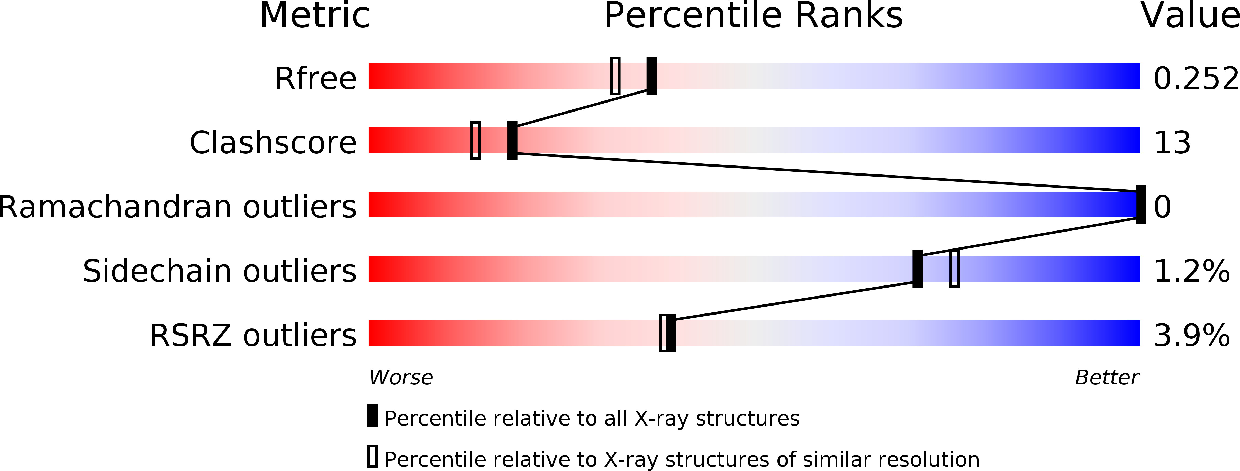
Resolution:
2.01 Å
R-Value Free:
0.23
R-Value Work:
0.17
R-Value Observed:
0.18
Space Group:
C 2 2 21
Macromolecular Entities
Polymer Type:polypeptide(L)
Molecule:Putative uncharacterized protein TB39.8
Gene (Uniprot):fhaA
Chain IDs:A
Chain Length:100
Number of Molecules:1
Biological Source:Mycobacterium tuberculosis
Polymer Type:polypeptide(L)
Molecule:synthetic phosphopeptide
Chain IDs:B
Chain Length:12
Number of Molecules:1
Biological Source:
Modified Residue
Compound ID Chain ID Parent Comp ID Details 2D Image
TPO B THR PHOSPHOTHREONINE
Ligand Molecules
Primary Citation
Structural and functional analysis of phosphothreonine-dependent FHA domain interactions
Structure 18 1587 1595 (2010)
PMID: 21134638 DOI: 10.1016/j.str.2010.09.014

Abstact

FHA domains are well established as phospho-dependent binding modules mediating signal transduction in Ser/Thr kinase signaling networks in both eukaryotic and prokaryotic species. Although they are unique in binding exclusively to phosphothreonine, the basis for this discrimination over phosphoserine has remained elusive. Here, we attempt to dissect overall binding specificity at the molecular level. We first determined the optimal peptide sequence for Rv0020c FHA domain binding by oriented peptide library screening. This served as a basis for systematic mutagenic and binding analyses, allowing us to derive relative thermodynamic contributions of conserved protein and peptide residues to binding and specificity. Structures of phosphopeptide-bound and uncomplexed Rv0020c FHA domain then directed molecular dynamics simulations which show how the extraordinary discrimination in favor of phosphothreonine occurs through formation of additional hydrogen-bonding networks that are ultimately stabilized by van der Waals interactions of the phosphothreonine γ-methyl group with a conserved pocket on the FHA domain surface.

Legend

Protein

Chemical

Disease

Primary Citation of related structures