3PNI image
Deposition Date 2010-11-19
Release Date 2011-04-13
Last Version Date 2024-11-06
Entry Detail
PDB ID:
3PNI
Title:
Crystal structure of D14C [3Fe-4S] Pyrococcus furiosus ferredoxin
Biological Source:
Source Organism:
Host Organism:
Method Details:
Experimental Method:
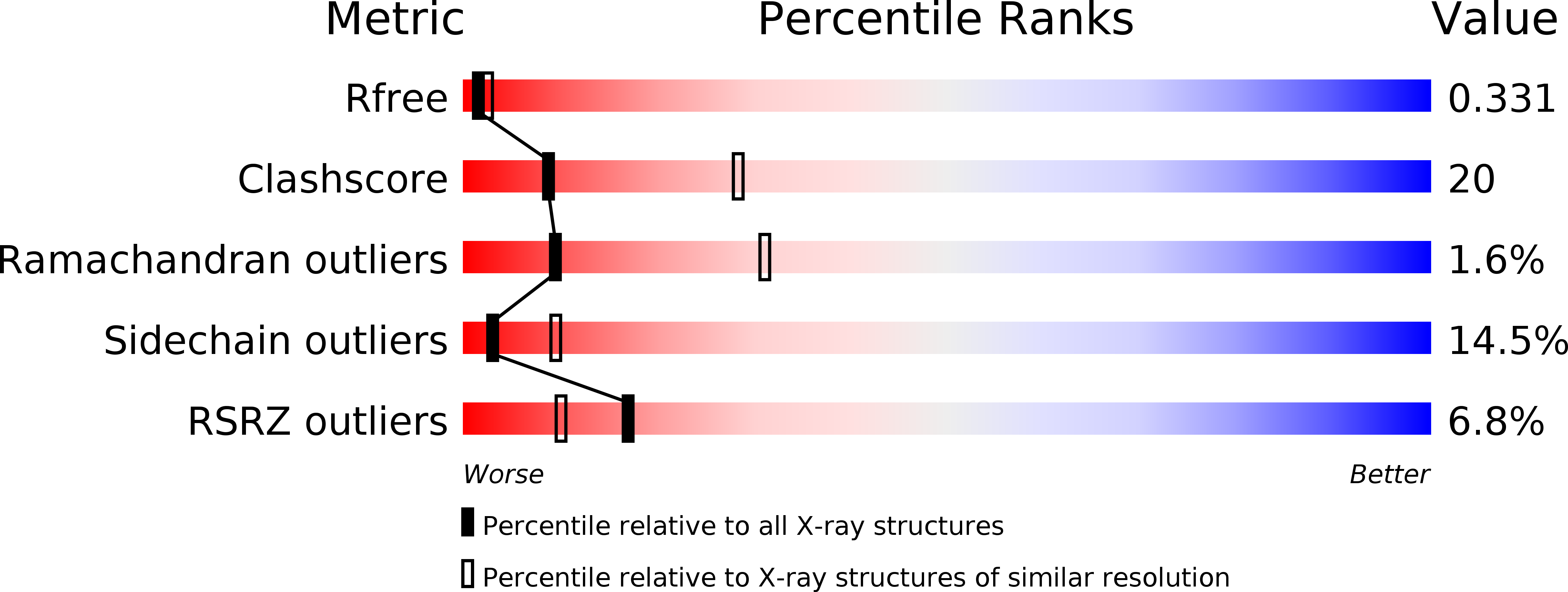
Resolution:
2.80 Å
R-Value Free:
0.31
R-Value Work:
0.27
R-Value Observed:
0.28
Space Group:
P 21 21 21
Macromolecular Entities
Polymer Type:polypeptide(L)
Molecule:Ferredoxin
Gene (Uniprot):fdxA
Mutations:D14C
Chain IDs:A, B
Chain Length:66
Number of Molecules:2
Biological Source:Pyrococcus furiosus
Primary Citation
Crystal structures of the all cysteinyl coordinated D14C variant of Pyrococcus furiosus ferredoxin: [4Fe-4S] <-> [3Fe-4S] cluster conversion
J.Biol.Inorg.Chem. 16 763 775 (2011)
PMID: 21484348 DOI: 10.1007/s00775-011-0778-7

Abstact

The structure of the all-cysteinyl-coordinated D14C variant of [4Fe-4S] ferredoxin from the hyperthermophilic archaeon Pyrococcus furiosus has been determined to 1.7 Å resolution from a crystal belonging to space group C222(1) with two types of molecules, A and B, in the asymmetric unit. A and B molecules have different crystal packing and intramolecular disulfide bond conformation. The crystal packing reveals a β-sheet interaction between A molecules in adjacent asymmetric units, whereas B molecules are packed as monomers in a less rigid position next to the A-A extended β-sheet dimers. The A molecules contain an intramolecular disulfide bond in a double conformation with 60% occupancy left-handed and 40% occupancy right-handed spiral conformation, whereas B molecules have an intramolecular disulfide bond in a right-handed spiral conformation. The cluster in D14C [4Fe-4S] P. furiosus ferredoxin was chemically oxidized at pH 5.8 to [3Fe-4S]. For purification at pH 8.0, two forms of the protein are obtained. Mass spectrometric analysis shows that the two forms are the D14C [3Fe-4S] P. furiosus ferredoxin monomer and a disulfide-bonded dimer of D14C [3Fe-4S] P. furiosus ferredoxin. When oxidization and purification are carried out at pH 5.8, only the monomer is obtained. The crystal structure of D14C [3Fe-4S] P. furiosus ferredoxin monomer was determined to 2.8 Å resolution from a crystal belonging to space group P2(1)2(1)2(1) with two molecules in the asymmetric unit. The molecules resemble molecule A of D14C [4Fe-4S] P. furiosus ferredoxin and electron density clearly shows the presence of a [3Fe-4S] cluster.

Legend

Protein

Chemical

Disease

Primary Citation of related structures