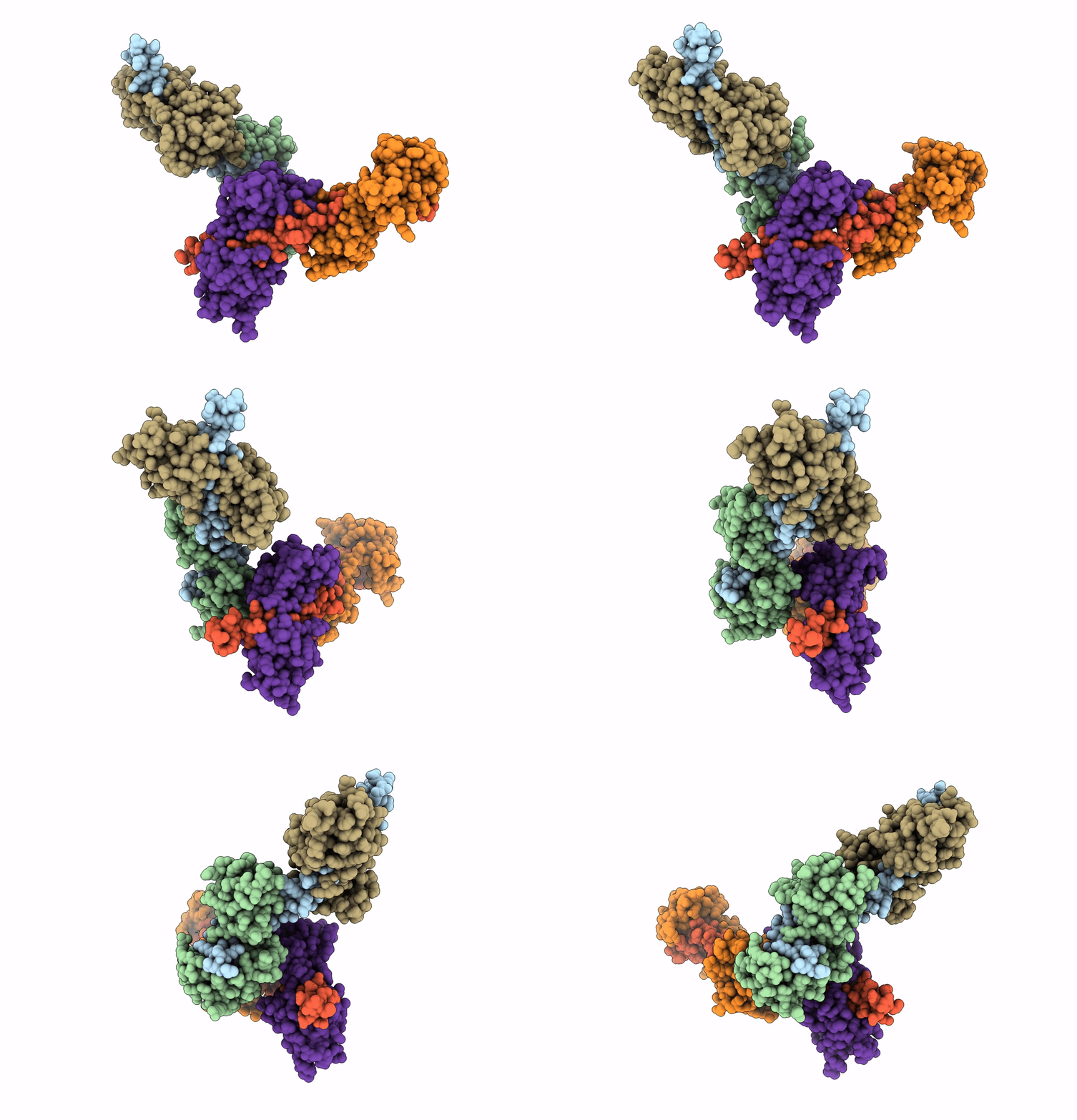
3PN7 image
Deposition Date 2010-11-18
Release Date 2011-01-05
Last Version Date 2023-09-06
Entry Detail
PDB ID:
3PN7
Keywords:
Title:
Visualizing new hinges and a potential major source of compliance in the lever arm of myosin
Biological Source:
Source Organism:
Method Details:
Experimental Method:
Resolution:
2.25 Å
R-Value Free:
0.24
R-Value Work:
0.19
R-Value Observed:
0.19
Space Group:
P 1
Macromolecular Entities
Structures with similar UniProt ID
Protein Blast
Polymer Type:polypeptide(L)
Molecule:Myosin heavy chain
Chain IDs:A, D
Chain Length:69
Number of Molecules:2
Biological Source:Placopecten magellanicus
Structures with similar UniProt ID
Protein Blast
Polymer Type:polypeptide(L)
Molecule:Myosin regulatory light chain
Chain IDs:B, E
Chain Length:161
Number of Molecules:2
Biological Source:Placopecten magellanicus
Structures with similar UniProt ID
Protein Blast
Polymer Type:polypeptide(L)
Molecule:Myosin essential light chain
Chain IDs:C, F
Chain Length:156
Number of Molecules:2
Biological Source:Placopecten magellanicus
Primary Citation
Visualizing key hinges and a potential major source of compliance in the lever arm of myosin.
Proc.Natl.Acad.Sci.USA 108 114 119 (2011)
PMID: 21149681 DOI: 10.1073/pnas.1016288107

Abstact

We have determined the 2.3-Å-resolution crystal structure of a myosin light chain domain, corresponding to one type found in sea scallop catch ("smooth") muscle. This structure reveals hinges that may function in the "on" and "off" states of myosin. The molecule adopts two different conformations about the heavy chain "hook" and regulatory light chain (RLC) helix D. This conformational change results in extended and compressed forms of the lever arm whose lengths differ by 10 Å. The heavy chain hook and RLC helix D hinges could thus serve as a potential major and localized source of cross-bridge compliance during the contractile cycle. In addition, in one of the molecules of the crystal, part of the RLC N-terminal extension is seen in atomic detail and forms a one-turn alpha-helix that interacts with RLC helix D. This extension, whose sequence is highly variable in different myosins, may thus modulate the flexibility of the lever arm. Moreover, the relative proximity of the phosphorylation site to the helix D hinge suggests a potential role for conformational changes about this hinge in the transition between the on and off states of regulated myosins.

Legend

Protein

Chemical

Disease

Primary Citation of related structures