3PMA image
Entry Detail
PDB ID:
3PMA
Title:
2.2 Angstrom crystal structure of the complex between Bovine Thrombin and Sucrose Octasulfate
Biological Source:
Source Organism:
PDB Version:
Deposition Date:
2010-11-16
Release Date:
2011-07-20
Method Details:
Experimental Method:
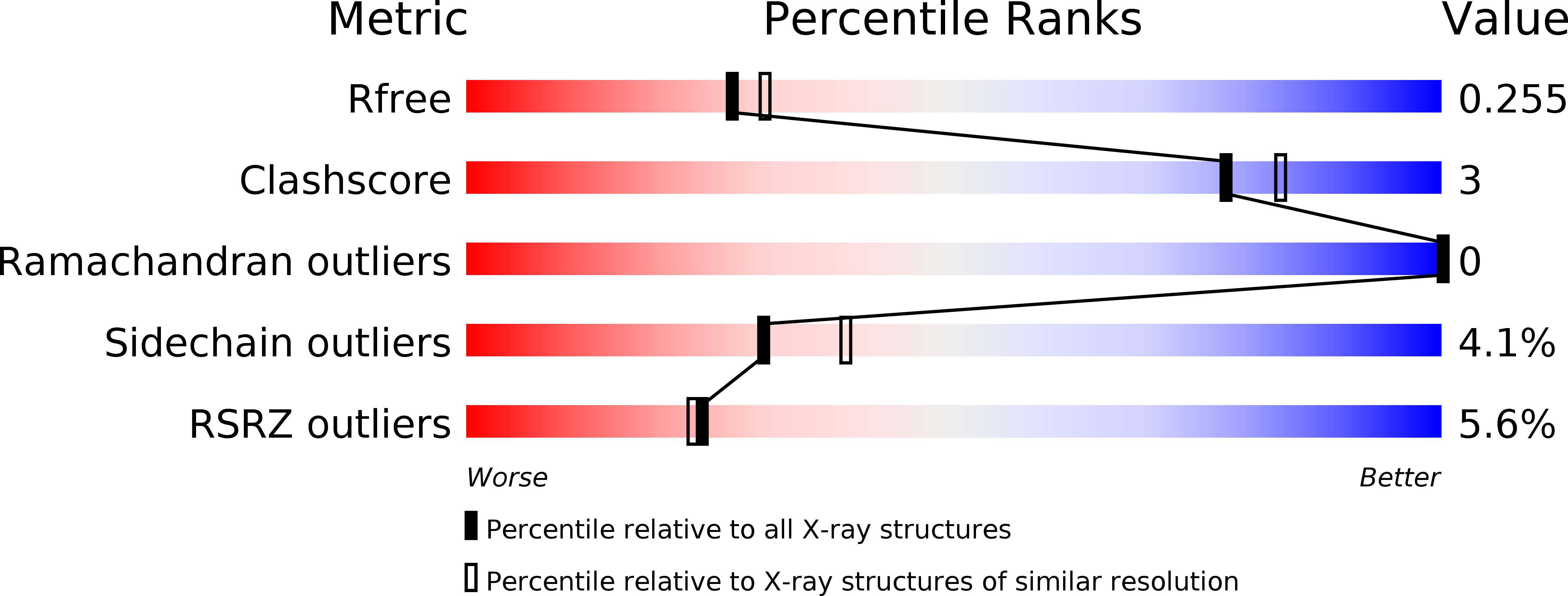
Resolution:
2.20 Å
R-Value Free:
0.24
R-Value Work:
0.20
R-Value Observed:
0.20
Space Group:
P 43 21 2
Macromolecular Entities
Polymer Type:polypeptide(L)
Description:Thrombin light chain
Chain IDs:A, C
Chain Length:29
Number of Molecules:2
Biological Source:Bos taurus
Polymer Type:polypeptide(L)
Description:Thrombin heavy chain
Chain IDs:B, D
Chain Length:259
Number of Molecules:2
Biological Source:Bos taurus
Peptide-like Molecules
PRD_900013
Primary Citation
Interaction of thrombin with sucrose octasulfate.
Biochemistry 50 6973 6982 (2011)
PMID: 21736375 DOI: 10.1021/bi2004526

Abstact

The serine protease thrombin plays multiple roles in many important physiological processes, especially coagulation, where it functions as both a pro- and anticoagulant. The polyanionic glycosaminoglycan heparin modulates thrombin's activity through binding at exosite II. Sucrose octasulfate (SOS) is often used as a surrogate for heparin, but it is not known whether it is an effective heparin mimic in its interaction with thrombin. We have characterized the interaction of SOS with thrombin in solution and determined a crystal structure of their complex. SOS binds thrombin with a K(d) of ~1.4 μM, comparable to that of the much larger polymeric heparin measured under the same conditions. Nonionic (hydrogen bonding) interactions make a larger contribution to thrombin binding of SOS than to heparin. SOS binding to exosite II inhibits thrombin's catalytic activity with high potency but with low efficacy. Analytical ultracentrifugation shows that bovine and human thrombins are monomers in solution in the presence of SOS, in contrast to their complexes with heparin, which are dimers. In the X-ray crystal structure, two molecules of SOS are bound nonequivalently to exosite II portions of a thrombin dimer, in contrast to the 1:2 stoichiometry of the heparin-thrombin complex, which has a different monomer association mode in the dimer. SOS and heparin binding to exosite II of thrombin differ on both chemical and structural levels and, perhaps most significantly, in thrombin inhibition. These differences may offer paths to the design of more potent exosite II binding, allosteric small molecules as modulators of thrombin function.

Legend

Protein

Chemical

Disease

Primary Citation of related structures