Deposition Date
2010-11-16
Release Date
2011-05-25
Last Version Date
2024-10-30
Entry Detail
PDB ID:
3PM2
Keywords:
Title:
Crystal structure of a novel type of odorant binding protein from Anopheles gambiae belonging to the c+ class
Biological Source:
Source Organism(s):
Anopheles gambiae (Taxon ID: 7165)
Expression System(s):
Method Details:
Experimental Method:
Resolution:
1.80 Å
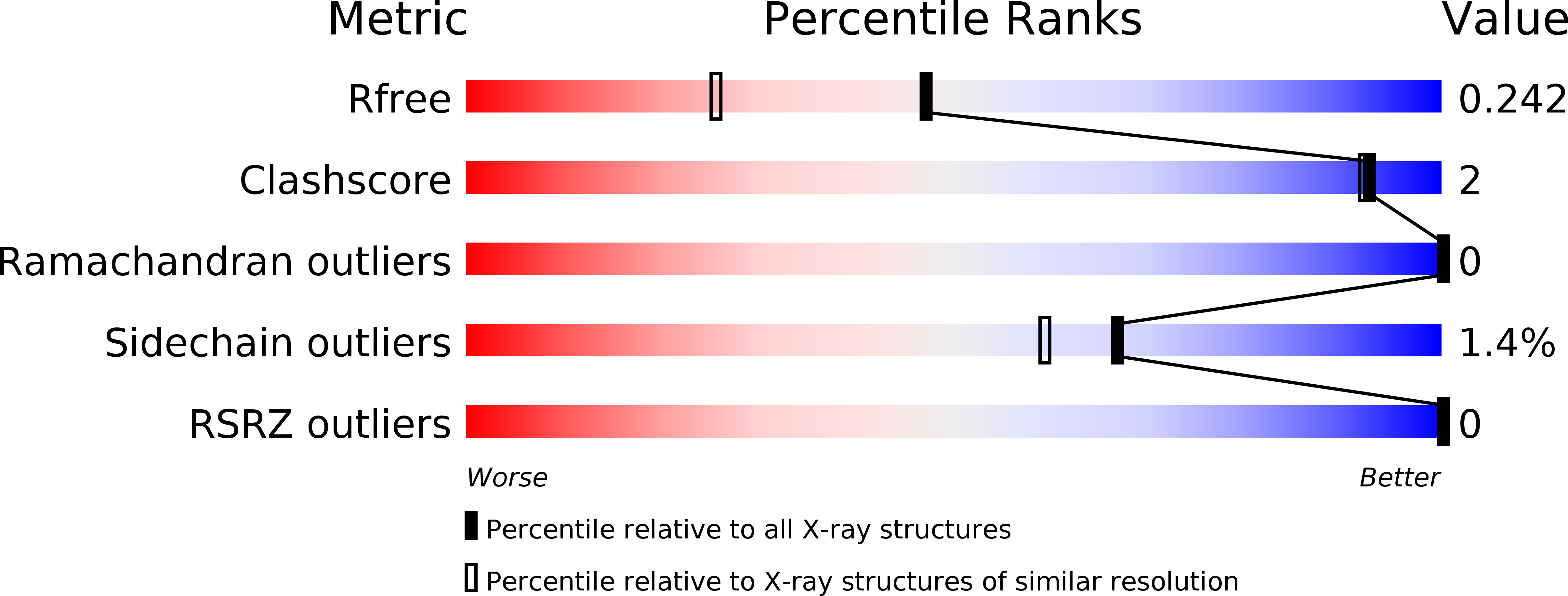
R-Value Free:
0.22
R-Value Work:
0.18
R-Value Observed:
0.18
Space Group:
P 41 21 2