3PL1 image
Deposition Date 2010-11-12
Release Date 2011-01-12
Last Version Date 2023-11-01
Entry Detail
PDB ID:
3PL1
Keywords:
Title:
Determination of the crystal structure of the pyrazinamidase from M.tuberculosis : a structure-function analysis for prediction resistance to pyrazinamide.
Biological Source:
Source Organism:
Host Organism:
Method Details:
Experimental Method:
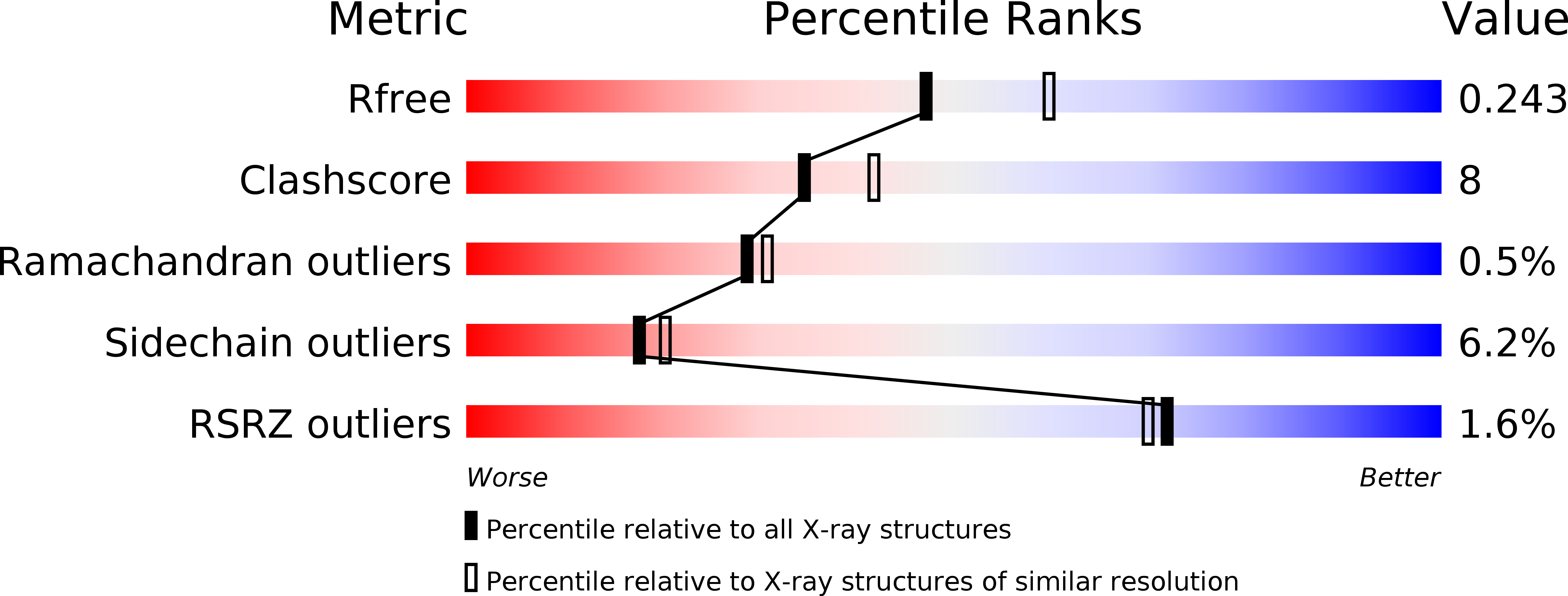
Resolution:
2.20 Å
R-Value Free:
0.23
R-Value Work:
0.19
R-Value Observed:
0.19
Space Group:
P 61 2 2
Macromolecular Entities
Polymer Type:polypeptide(L)
Molecule:PYRAZINAMIDASE/NICOTINAMIDASE PNCA (PZase)
Chain IDs:A
Chain Length:186
Number of Molecules:1
Biological Source:Mycobacterium tuberculosis
Ligand Molecules
Primary Citation
Crystal Structure of the Pyrazinamidase of Mycobacterium tuberculosis: Insights into Natural and Acquired Resistance to Pyrazinamide.
Plos One 6 e15785 e15785 (2011)
PMID: 21283666 DOI: 10.1371/journal.pone.0015785

Abstact

Pyrazinamidase (PncA) activates the first-line antituberculous drug pyrazinamide into pyrazinoic acid. The crystal structure of the Mycobacterium tuberculosis PncA protein has been determined, showing significant differences in the substrate binding cavity when compared to the pyrazinamidases from Pyrococcus horikoshii and Acinetobacter baumanii. In M. tuberculosis, this region was found to hold a Fe(2+) ion coordinated by one aspartate and three histidines, one of them corresponding to His57 which is replaced by Asp in Mycobacterium bovis, a species naturally resistant to pyrazinamide. The binding cavity also contains a Cys138-Asp8-Lys96 motif evocating a cysteine-based catalytic mechanism. Mutants have been constructed and investigated by kinetic and thermal shift assays, highlighting the importance of protein folding and thermal stability in the pyrazinamidase activity.

Legend

Protein

Chemical

Disease

Primary Citation of related structures