3PKY image
Deposition Date 2010-11-12
Release Date 2011-02-16
Last Version Date 2024-02-21
Entry Detail
PDB ID:
3PKY
Keywords:
Title:
Polymerase Domain from Mycobacterium tuberculosis Ligase D in complex with DNA, UTP and Manganese.
Biological Source:
Source Organism:
Host Organism:
Method Details:
Experimental Method:
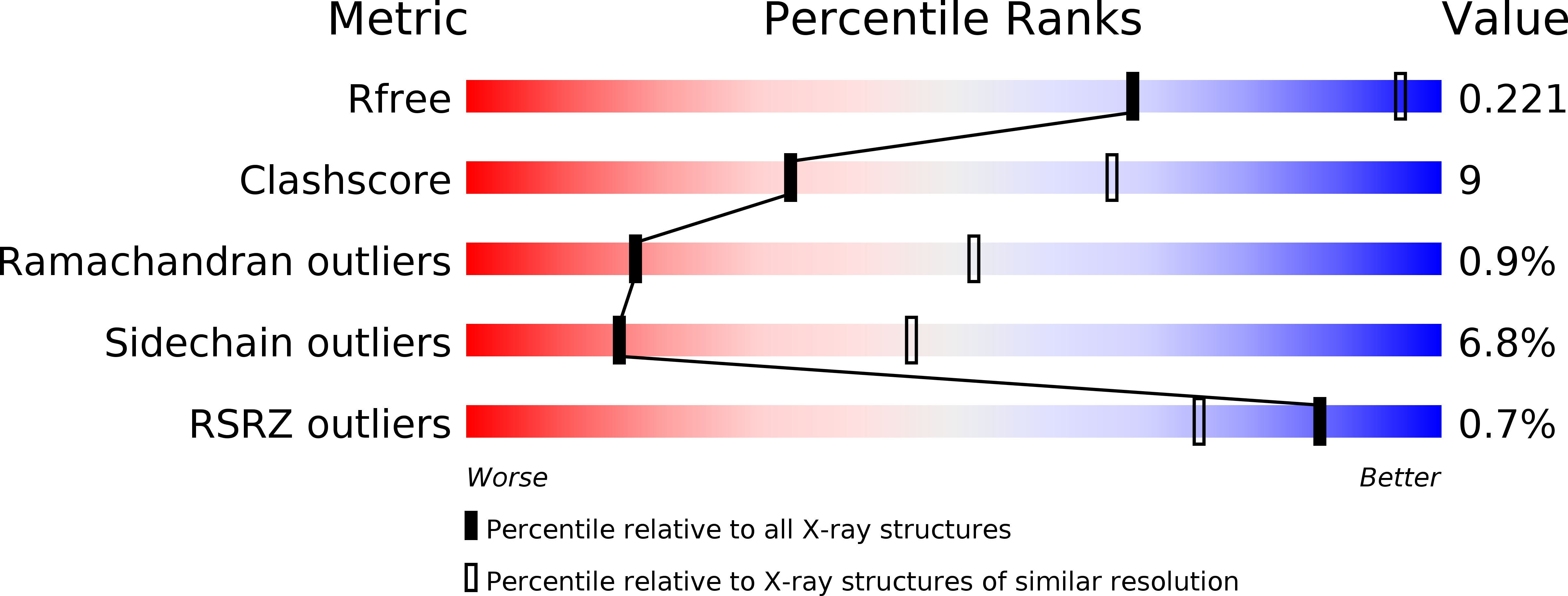
Resolution:
3.10 Å
R-Value Free:
0.24
R-Value Work:
0.19
R-Value Observed:
0.19
Space Group:
P 41
Macromolecular Entities
Polymer Type:polypeptide(L)
Molecule:Putative DNA ligase-like protein
Chain IDs:A, B
Chain Length:303
Number of Molecules:2
Biological Source:Mycobacterium tuberculosis
Primary Citation
Structure of a Preternary Complex Involving a Prokaryotic NHEJ DNA Polymerase.
Mol.Cell 41 221 231 (2011)
PMID: 21255731 DOI: 10.1016/j.molcel.2010.12.026

Abstact

In many prokaryotes, a specific DNA primase/polymerase (PolDom) is required for nonhomologous end joining (NHEJ) repair of DNA double-strand breaks (DSBs). Here, we report the crystal structure of a catalytically active conformation of Mycobacterium tuberculosis PolDom, consisting of a polymerase bound to a DNA end with a 3' overhang, two metal ions, and an incoming nucleotide but, significantly, lacking a primer strand. This structure represents a polymerase:DNA complex in a preternary intermediate state. This polymerase complex occurs in solution, stabilizing the enzyme on DNA ends and promoting nucleotide extension of short incoming termini. We also demonstrate that the invariant Arg(220), contained in a conserved loop (loop 2), plays an essential role in catalysis by regulating binding of a second metal ion in the active site. We propose that this NHEJ intermediate facilitates extension reactions involving critically short or noncomplementary DNA ends, thus promoting break repair and minimizing sequence loss during DSB repair.

Legend

Protein

Chemical

Disease

Primary Citation of related structures