
Deposition Date
2010-11-10
Release Date
2011-09-28
Last Version Date
2023-11-01
Entry Detail
PDB ID:
3PJG
Keywords:
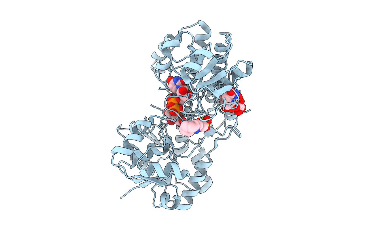
Title:
Crystal structure of UDP-glucose dehydrogenase from Klebsiella pneumoniae complexed with product UDP-glucuronic acid
Biological Source:
Source Organism(s):
Klebsiella pneumoniae (Taxon ID: 573)
Expression System(s):
Method Details:
Experimental Method:
Resolution:
2.70 Å
R-Value Free:
0.22
R-Value Work:
0.18
R-Value Observed:
0.18
Space Group:
P 64 2 2
