Deposition Date
2010-11-07
Release Date
2011-02-23
Last Version Date
2024-10-09
Entry Detail
PDB ID:
3PIP
Keywords:
Title:
Crystal structure of the synergistic antibiotic pair lankamycin and lankacidin in complex with the large ribosomal subunit
Biological Source:
Source Organism(s):
Deinococcus radiodurans R1 (Taxon ID: 243230)
Deinococcus radiodurans (Taxon ID: 1299)
Deinococcus radiodurans (Taxon ID: 1299)
Method Details:
Experimental Method:
Resolution:
3.45 Å
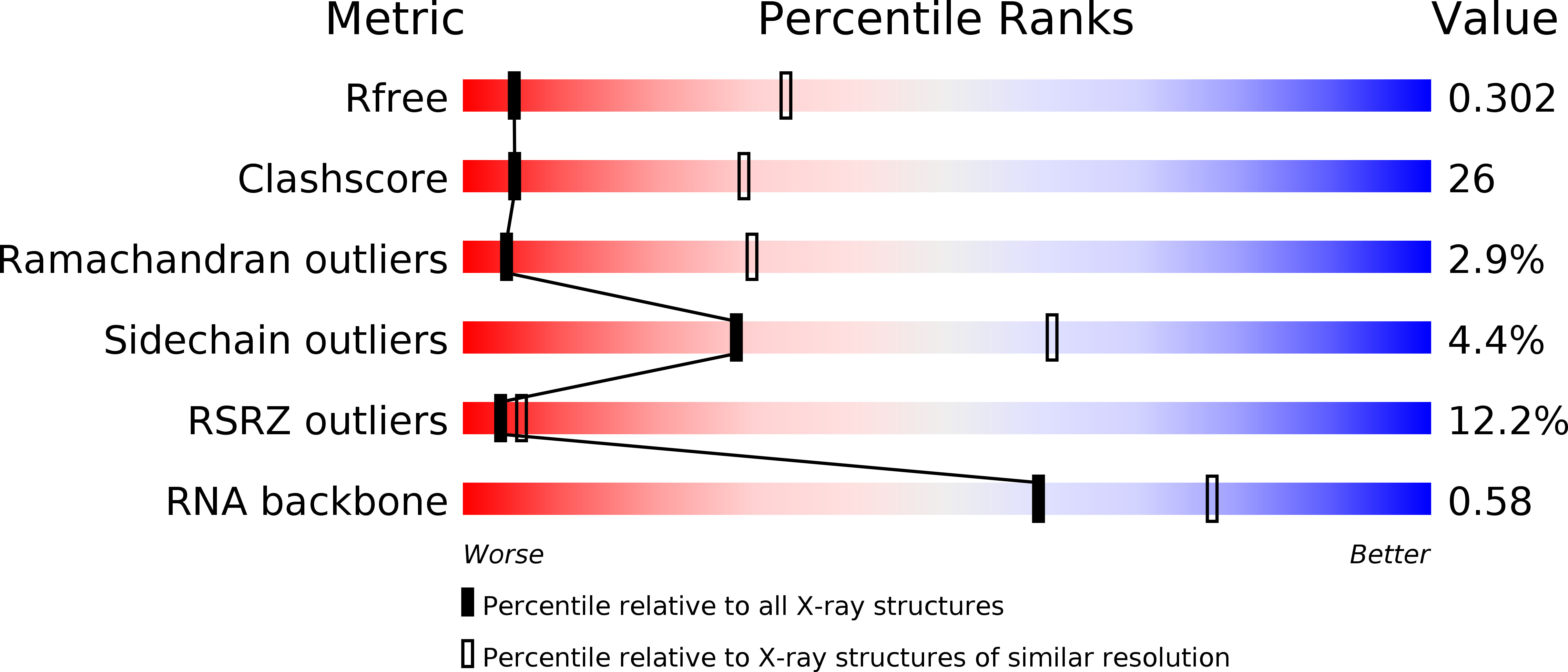
R-Value Free:
0.30
R-Value Work:
0.25
R-Value Observed:
0.25
Space Group:
I 2 2 2