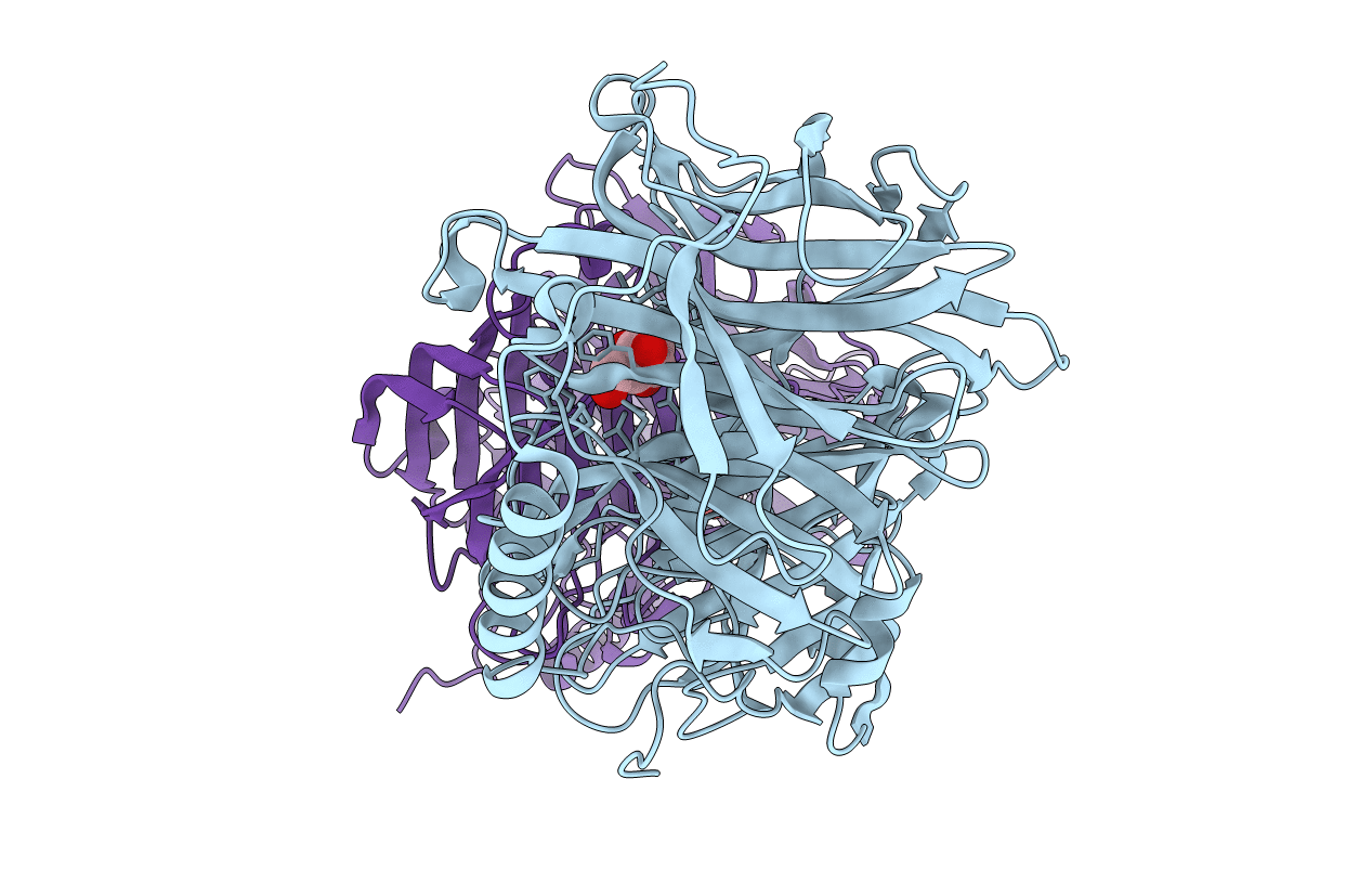
3PIJ image
Deposition Date 2010-11-06
Release Date 2011-04-27
Last Version Date 2023-09-06
Entry Detail
PDB ID:
3PIJ
Keywords:
Title:
beta-fructofuranosidase from Bifidobacterium longum - complex with fructose
Biological Source:
Source Organism:
Host Organism:
Method Details:
Experimental Method:
Resolution:
1.80 Å
R-Value Free:
0.20
R-Value Work:
0.15
R-Value Observed:
0.15
Space Group:
P 31 2 1
Macromolecular Entities
Polymer Type:polypeptide(L)
Molecule:Beta-fructofuranosidase
Gene (Uniprot):B5U78_03730, CE169_09530, DWV23_02165, DWV59_08965, DXC63_04880, DXC85_10215, GBC97_07305, GBI83_00975, GBI87_04425, GBK08_07415, KSW34_10515
Chain IDs:A, B
Chain Length:526
Number of Molecules:2
Biological Source:Bifidobacterium longum
Primary Citation
Crystal structures of the apo form of beta-fructofuranosidase from Bifidobacterium longum and its complex with fructose
Febs J. 278 1728 1744 (2011)
PMID: 21418142 DOI: 10.1111/j.1742-4658.2011.08098.x

Abstact

We solved the 1.8 Å crystal structure of β-fructofuranosidase from Bifidobacterium longum KN29.1 - a unique enzyme that allows these probiotic bacteria to function in the human digestive system. The sequence of β-fructofuranosidase classifies it as belonging to the glycoside hydrolase family 32 (GH32). GH32 enzymes show a wide range of substrate specificity and different functions in various organisms. All enzymes from this family share a similar fold, containing two domains: an N-terminal five-bladed β-propeller and a C-terminal β-sandwich module. The active site is located in the centre of the β-propeller domain, in the bottom of a 'funnel'. The binding site, -1, responsible for tight fructose binding, is highly conserved among the GH32 enzymes. Bifidobacterium longum KN29.1 β-fructofuranosidase has a 35-residue elongation of the N-terminus containing a five-turn α-helix, which distinguishes it from the other known members of the GH32 family. This new structural element could be one of the functional modifications of the enzyme that allows the bacteria to act in a human digestive system. We also solved the 1.8 Å crystal structure of the β-fructofuranosidase complex with β-D-fructose, a hydrolysis product obtained by soaking apo crystal in raffinose.

Legend

Protein

Chemical

Disease

Primary Citation of related structures