3PIH image
Deposition Date 2010-11-06
Release Date 2011-01-19
Last Version Date 2023-09-06
Entry Detail
PDB ID:
3PIH
Keywords:
Title:
T. maritima UvrA in complex with fluorescein-modified DNA
Biological Source:
Source Organism:
Host Organism:
Method Details:
Experimental Method:
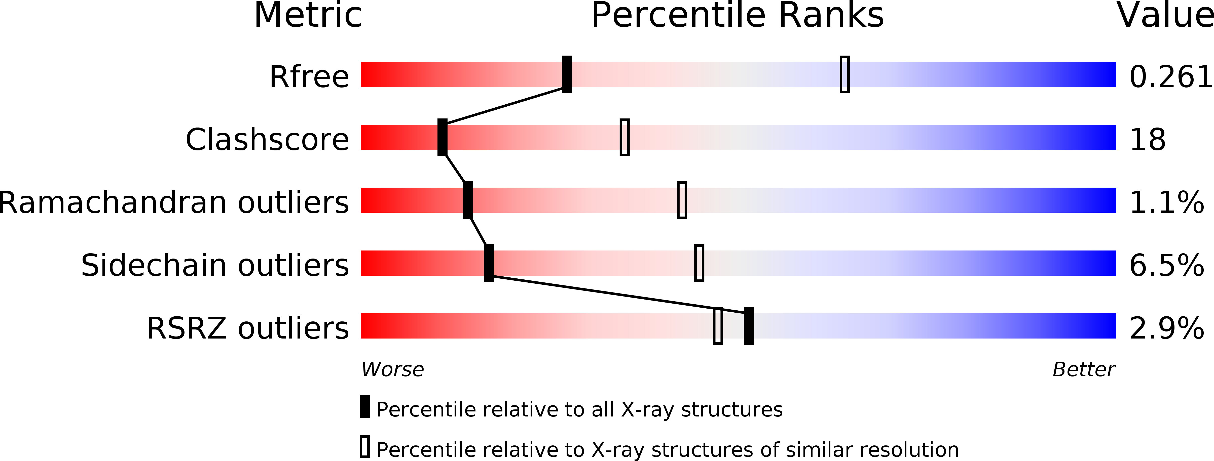
Resolution:
2.90 Å
R-Value Free:
0.26
R-Value Work:
0.19
R-Value Observed:
0.20
Space Group:
P 42
Macromolecular Entities
Structures with similar UniProt ID
Protein Blast
Polymer Type:polypeptide(L)
Molecule:UvrABC system protein A
Gene (Uniprot):uvrA
Chain IDs:A
Chain Length:916
Number of Molecules:1
Biological Source:Thermotoga maritima
Polymer Type:polydeoxyribonucleotide
Molecule:DNA (32-MER)
Chain IDs:B (auth: D)
Chain Length:32
Number of Molecules:1
Biological Source:
Primary Citation
Structure of UvrA nucleotide excision repair protein in complex with modified DNA.
Nat.Struct.Mol.Biol. 18 191 197 (2011)
PMID: 21240268 DOI: 10.1038/nsmb.1973

Abstact

One of the primary pathways for removal of DNA damage is nucleotide excision repair (NER). In bacteria, the UvrA protein is the component of NER that locates the lesion. A notable feature of NER is its ability to act on many DNA modifications that vary in chemical structure. So far, the mechanism underlying this broad specificity has been unclear. Here, we report the first crystal structure of a UvrA protein in complex with a chemically modified oligonucleotide. The structure shows that the UvrA dimer does not contact the site of lesion directly, but rather binds the DNA regions on both sides of the modification. The DNA region harboring the modification is deformed, with the double helix bent and unwound. UvrA uses damage-induced deformations of the DNA and a less rigid structure of the modified double helix for indirect readout of the lesion.

Legend

Protein

Chemical

Disease

Primary Citation of related structures
Feedback Form
Name
Email
Institute
Feedback