3PIF image
Deposition Date 2010-11-06
Release Date 2011-02-09
Last Version Date 2024-02-21
Entry Detail
PDB ID:
3PIF
Keywords:
Title:
Crystal structure of the 5'->3' exoribonuclease Xrn1, E178Q mutant in Complex with Manganese
Biological Source:
Source Organism:
Host Organism:
Method Details:
Experimental Method:
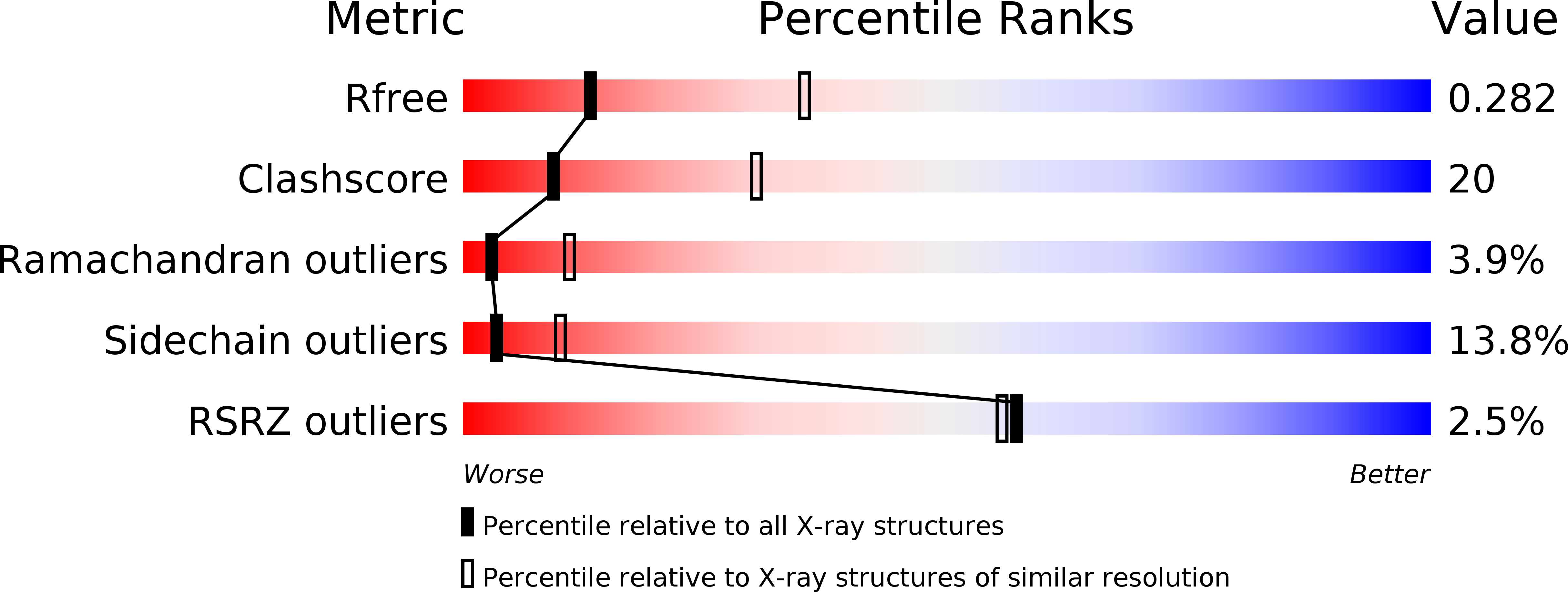
Resolution:
2.92 Å
R-Value Free:
0.28
R-Value Work:
0.24
R-Value Observed:
0.24
Space Group:
P 1
Macromolecular Entities
Structures with similar UniProt ID
Protein Blast
Polymer Type:polypeptide(L)
Molecule:5'->3' EXORIBONUCLEASE (xrn1)
Gene (Uniprot):KLLA0_F22385g
Mutagens:E178Q
Chain IDs:A, B, C, D
Chain Length:1155
Number of Molecules:4
Biological Source:Kluyveromyces lactis
Ligand Molecules
Primary Citation
Structural and biochemical studies of the 5' -> 3' exoribonuclease Xrn1.
Nat.Struct.Mol.Biol. 18 270 276 (2011)
PMID: 21297639 DOI: 10.1038/nsmb.1984

Abstact

The 5'→3' exoribonucleases (XRNs) have important functions in transcription, RNA metabolism and RNA interference. The structure of Rat1 (also known as Xrn2) showed that the two highly conserved regions of XRNs form a single, large domain that defines the active site of the enzyme. Xrn1 has a 510-residue segment after the conserved regions that is required for activity but is absent from Rat1/Xrn2. Here we report the crystal structures of Kluyveromyces lactis Xrn1 (residues 1-1,245, E178Q mutant), alone and in complex with a Mn(2+) ion in the active site. The 510-residue segment contains four domains (D1-D4), located far from the active site. Our mutagenesis and biochemical studies show that their functional importance results from their ability to stabilize the conformation of the N-terminal segment of Xrn1. These domains might also constitute a platform that interacts with protein partners of Xrn1.

Legend

Protein

Chemical

Disease

Primary Citation of related structures