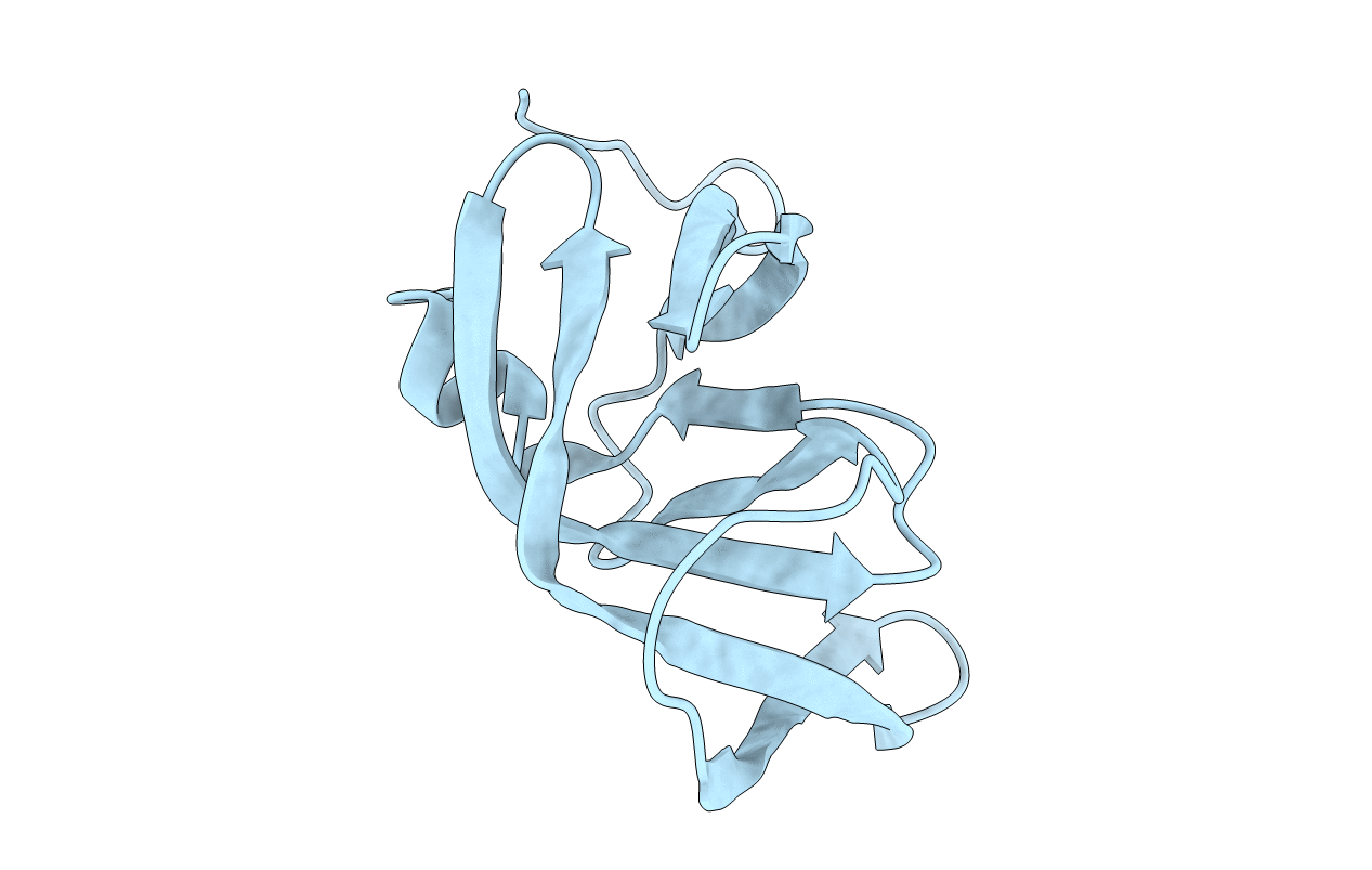
3PHV image
Deposition Date 1991-11-04
Release Date 1992-01-15
Last Version Date 2024-02-21
Entry Detail
PDB ID:
3PHV
Keywords:
Title:
X-RAY ANALYSIS OF HIV-1 PROTEINASE AT 2.7 ANGSTROMS RESOLUTION CONFIRMS STRUCTURAL HOMOLOGY AMONG RETROVIRAL ENZYMES
Biological Source:
Source Organism:
Host Organism:
Method Details:
Experimental Method:
Resolution:
2.70 Å
R-Value Work:
0.19
Space Group:
P 41 21 2
Macromolecular Entities
Structures with similar UniProt ID
Protein Blast
Polymer Type:polypeptide(L)
Molecule:UNLIGANDED HIV-1 PROTEASE
Gene (Uniprot):gag-pol
Chain IDs:A
Chain Length:99
Number of Molecules:1
Biological Source:HIV-1 M:B_HXB2R
Ligand Molecules
Primary Citation
X-ray analysis of HIV-1 proteinase at 2.7 A resolution confirms structural homology among retroviral enzymes.
Nature 342 299 302 (1989)
PMID: 2682266 DOI: 10.1038/342299a0

Abstact

Knowledge of the tertiary structure of the proteinase from human immunodeficiency virus HIV-1 is important to the design of inhibitors that might possess antiviral activity and thus be useful in the treatment of AIDS. The conserved Asp-Thr/Ser-Gly sequence in retroviral proteinases suggests that they exist as dimers similar to the ancestor proposed for the pepsins. Although this has been confirmed by X-ray analyses of Rous sarcoma virus and HIV-1 proteinases, these structures have overall folds that are similar to each other only where they are also similar to the pepsins. We now report a further X-ray analysis of a recombinant HIV-1 proteinase at 2.7 A resolution. The polypeptide chain adopts a fold in which the N- and C-terminal strands are organized together in a four-stranded beta-sheet. A helix precedes the single C-terminal strand, as in the Rous sarcoma virus proteinase and also in a synthetic HIV-1 proteinase, in which the cysteines have been replaced by alpha-aminobuytric acid. The structure reported here provides an explanation for the amino acid invariance amongst retroviral proteinases, but differs from that reported earlier in some residues that are candidates for substrate interactions at P3, and in the mode of intramolecular cleavage during processing of the polyprotein.

Legend

Protein

Chemical

Disease

Primary Citation of related structures