3PH0 image
Deposition Date 2010-11-03
Release Date 2011-07-20
Last Version Date 2024-03-20
Entry Detail
PDB ID:
3PH0
Keywords:
Title:
Crystal structure of the heteromolecular chaperone, AscE-AscG, from the type III secretion system in Aeromonas hydrophila
Biological Source:
Source Organism:
Host Organism:
Method Details:
Experimental Method:
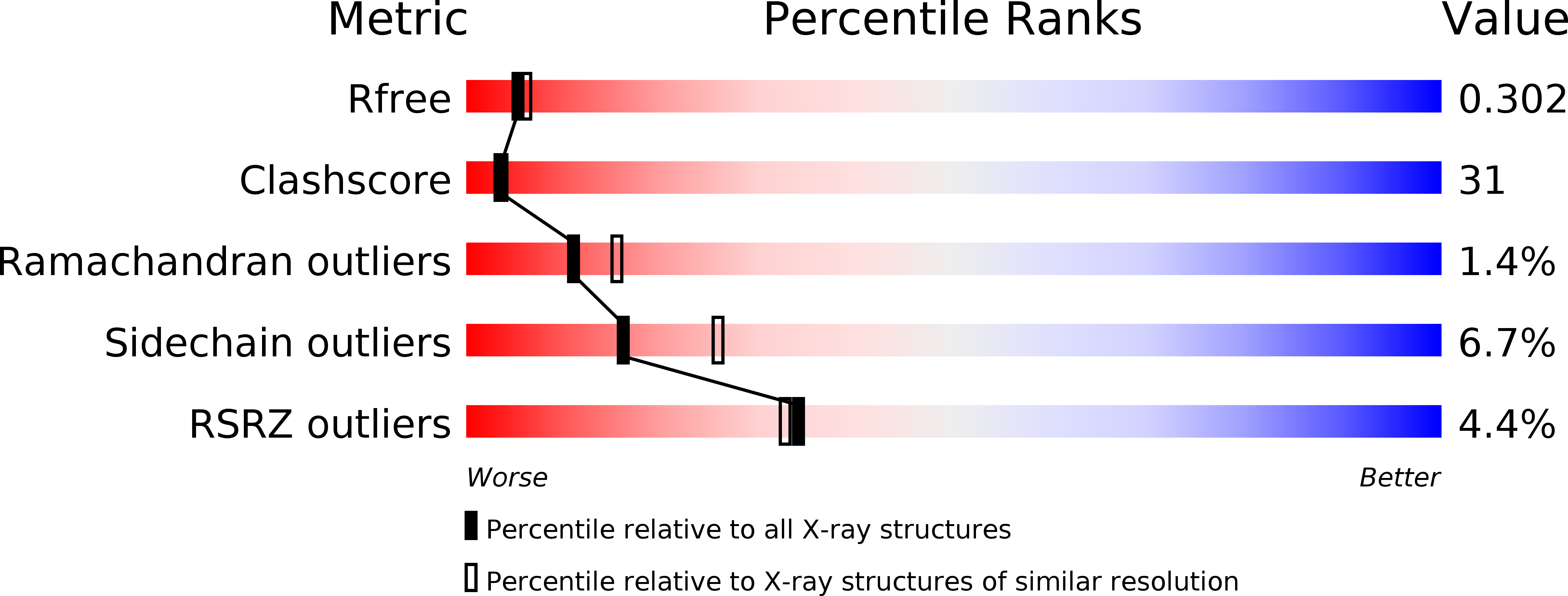
Resolution:
2.40 Å
R-Value Free:
0.29
R-Value Work:
0.23
Space Group:
P 21 21 2
Macromolecular Entities
Polymer Type:polypeptide(L)
Molecule:AscE
Gene (Uniprot):ascE
Chain IDs:A, C (auth: B)
Chain Length:67
Number of Molecules:2
Biological Source:Aeromonas hydrophila
Polymer Type:polypeptide(L)
Molecule:AscG
Gene (Uniprot):ascG
Chain IDs:B (auth: C), D
Chain Length:61
Number of Molecules:2
Biological Source:Aeromonas hydrophila
Primary Citation
Crystal structure of the heteromolecular chaperone, AscE-AscG, from the type III secretion system in Aeromonas hydrophila
Plos One 6 e19208 e19208 (2011)
PMID: 21559439 DOI: 10.1371/journal.pone.0019208

Abstact

BACKGROUND The putative needle complex subunit AscF forms a ternary complex with the chaperones AscE and AscG in the type III secretion system of Aeromonas hydrophila so as to avoid premature assembly. Previously, we demonstrated that the C-terminal region of AscG (residues 62-116) in the hetero-molecular chaperone, AscE-AscG, is disordered and susceptible to limited protease digestion. METHODOLOGY/PRINCIPAL FINDINGS Here, we report the crystal structure of the ordered AscG(1-61) region in complex with AscE at 2.4 Å resolution. Helices α2 and α3 of AscE in the AscE-AscG(1-61) complex assumes a helix-turn-helix conformation in an anti-parallel fashion similar to that in apo AscE. However, in the presence of AscG, an additional N-terminal helix α1 in AscE (residues 4-12) is observed. PscG or YscG in the crystal structures of PscE-PscF-PscG or YscE-YscF-YscG, respectively, assumes a typical tetratricopeptide repeat (TPR) fold with three TPR repeats and one C-terminal capping helix. By comparison, AscG in AscE-AscG(1-61) comprises three anti-parallel helices that resembles the N-terminal TPR repeats in the corresponding region of PscG or YscG in PscE-PscF-PscG or YscE-YscF-YscG. Thermal denaturation of AscE-AscG and AscE-AscG(1-61) complexes demonstrates that the C-terminal disordered region does not contribute to the thermal stability of the overall complex. CONCLUSION/SIGNIFICANCE The N-terminal region of the AscG in the AscE-AscG complex is ordered and assumes a structure similar to those in the corresponding regions of PscE-PscG-PscF or YscE-YscF-YscG complexes. While the C-terminal region of AscG in the AscE-AscG complex is disordered and will assume its structure only in the presence of the substrate AscF. We hypothesize that AscE act as a chaperone of the chaperone to keep AscG in a stable but partially disordered state for interaction with AscF.

Legend

Protein

Chemical

Disease

Primary Citation of related structures