Deposition Date
2010-10-29
Release Date
2011-03-02
Last Version Date
2024-11-20
Entry Detail
PDB ID:
3PG2
Keywords:
Title:
The Crystal structure of the major pilin GBS80 of Streptococcus agalactiae 35 kDa C-terminal fragment
Biological Source:
Source Organism(s):
Streptococcus agalactiae serogroup V (Taxon ID: 216466)
Expression System(s):
Method Details:
Experimental Method:
Resolution:
1.80 Å
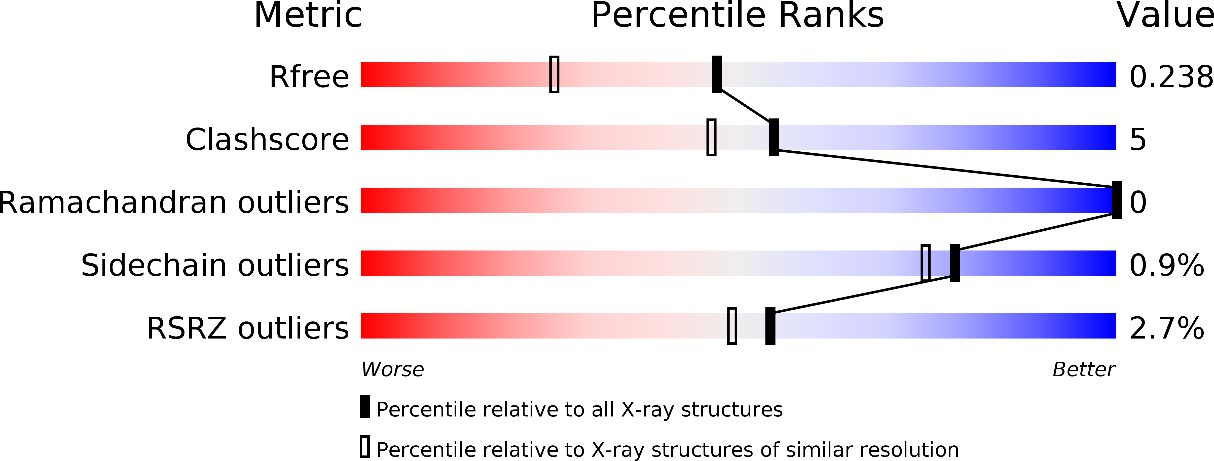
R-Value Free:
0.23
R-Value Work:
0.20
R-Value Observed:
0.20
Space Group:
P 1 21 1