3PE4 image
Deposition Date 2010-10-25
Release Date 2011-01-19
Last Version Date 2024-02-21
Entry Detail
PDB ID:
3PE4
Keywords:
Title:
Structure of human O-GlcNAc transferase and its complex with a peptide substrate
Biological Source:
Source Organism:
Homo sapiens (Taxon ID: 9606)
Host Organism:
Method Details:
Experimental Method:
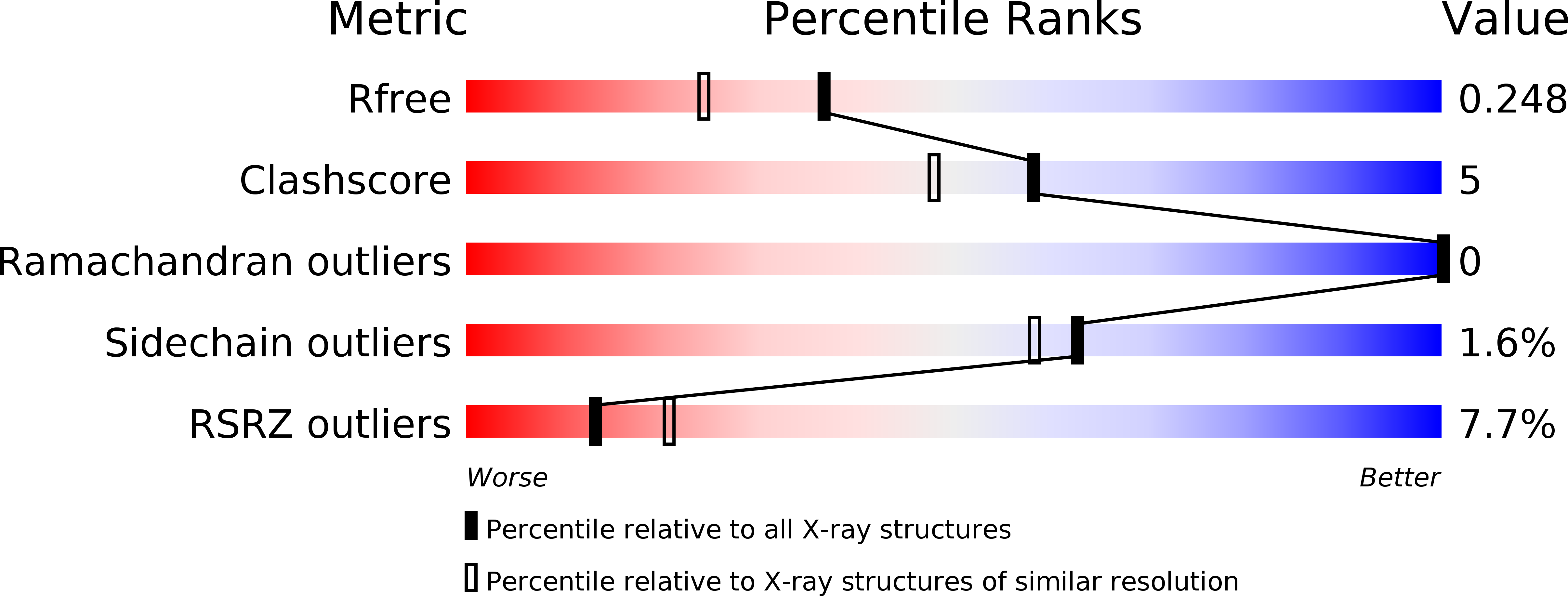
Resolution:
1.95 Å
R-Value Free:
0.25
R-Value Work:
0.22
R-Value Observed:
0.22
Space Group:
I 1 2 1
Macromolecular Entities
Structures with similar UniProt ID
Protein Blast
Polymer Type:polypeptide(L)
Molecule:UDP-N-acetylglucosamine--peptide N-acetylglucosaminyltransferase 110 kDa subunit
Gene (Uniprot):OGT
Chain IDs:A, C
Chain Length:723
Number of Molecules:2
Biological Source:Homo sapiens
Structures with similar UniProt ID
Protein Blast
Polymer Type:polypeptide(L)
Molecule:Casein kinase II subunit alpha
Gene (Uniprot):CSNK2A1
Chain IDs:B, D
Chain Length:14
Number of Molecules:2
Biological Source:Homo sapiens
Primary Citation
Structure of human O-GlcNAc transferase and its complex with a peptide substrate.
Nature 469 564 567 (2011)
PMID: 21240259 DOI: 10.1038/nature09638

Abstact

The essential mammalian enzyme O-linked β-N-acetylglucosamine transferase (O-GlcNAc transferase, here OGT) couples metabolic status to the regulation of a wide variety of cellular signalling pathways by acting as a nutrient sensor. OGT catalyses the transfer of N-acetylglucosamine from UDP-N-acetylglucosamine (UDP-GlcNAc) to serines and threonines of cytoplasmic, nuclear and mitochondrial proteins, including numerous transcription factors, tumour suppressors, kinases, phosphatases and histone-modifying proteins. Aberrant glycosylation by OGT has been linked to insulin resistance, diabetic complications, cancer and neurodegenerative diseases including Alzheimer's. Despite the importance of OGT, the details of how it recognizes and glycosylates its protein substrates are largely unknown. We report here two crystal structures of human OGT, as a binary complex with UDP (2.8 Å resolution) and as a ternary complex with UDP and a peptide substrate (1.95 Å). The structures provide clues to the enzyme mechanism, show how OGT recognizes target peptide sequences, and reveal the fold of the unique domain between the two halves of the catalytic region. This information will accelerate the rational design of biological experiments to investigate OGT's functions; it will also help the design of inhibitors for use as cellular probes and help to assess its potential as a therapeutic target.

Legend

Protein

Chemical

Disease

Primary Citation of related structures
Feedback Form
Name
Email
Institute
Feedback