
Deposition Date
2010-10-24
Release Date
2011-01-12
Last Version Date
2024-10-30
Entry Detail
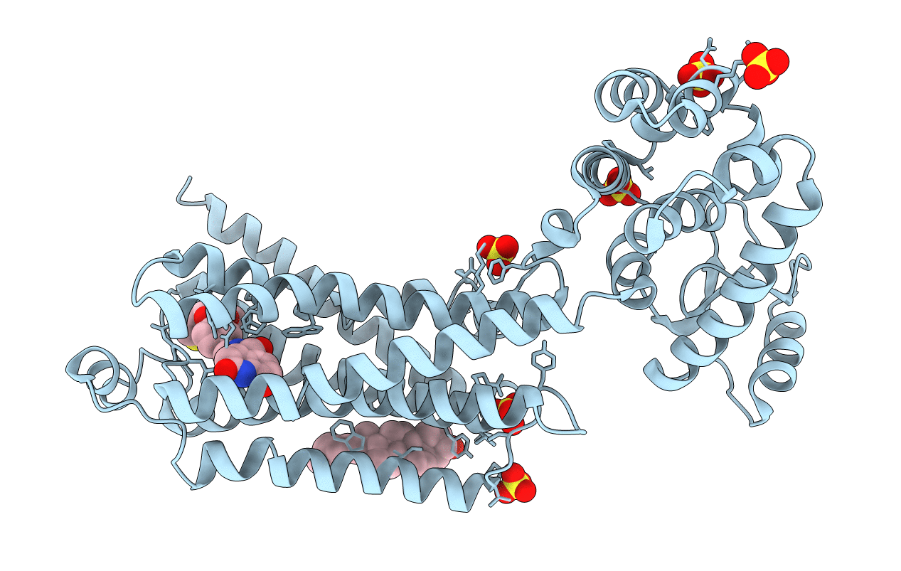
PDB ID:
3PDS
Keywords:
Title:
Irreversible Agonist-Beta2 Adrenoceptor Complex
Biological Source:
Source Organism(s):
Homo sapiens (Taxon ID: 9606)
Enterobacteria phage T4 (Taxon ID: 10665)
Enterobacteria phage T4 (Taxon ID: 10665)
Expression System(s):
Method Details:
Experimental Method:
Resolution:
3.50 Å
R-Value Free:
0.28
R-Value Work:
0.23
R-Value Observed:
0.24
Space Group:
P 21 21 2
