3PD8 image
Deposition Date 2010-10-22
Release Date 2010-12-29
Last Version Date 2024-11-27
Entry Detail
PDB ID:
3PD8
Title:
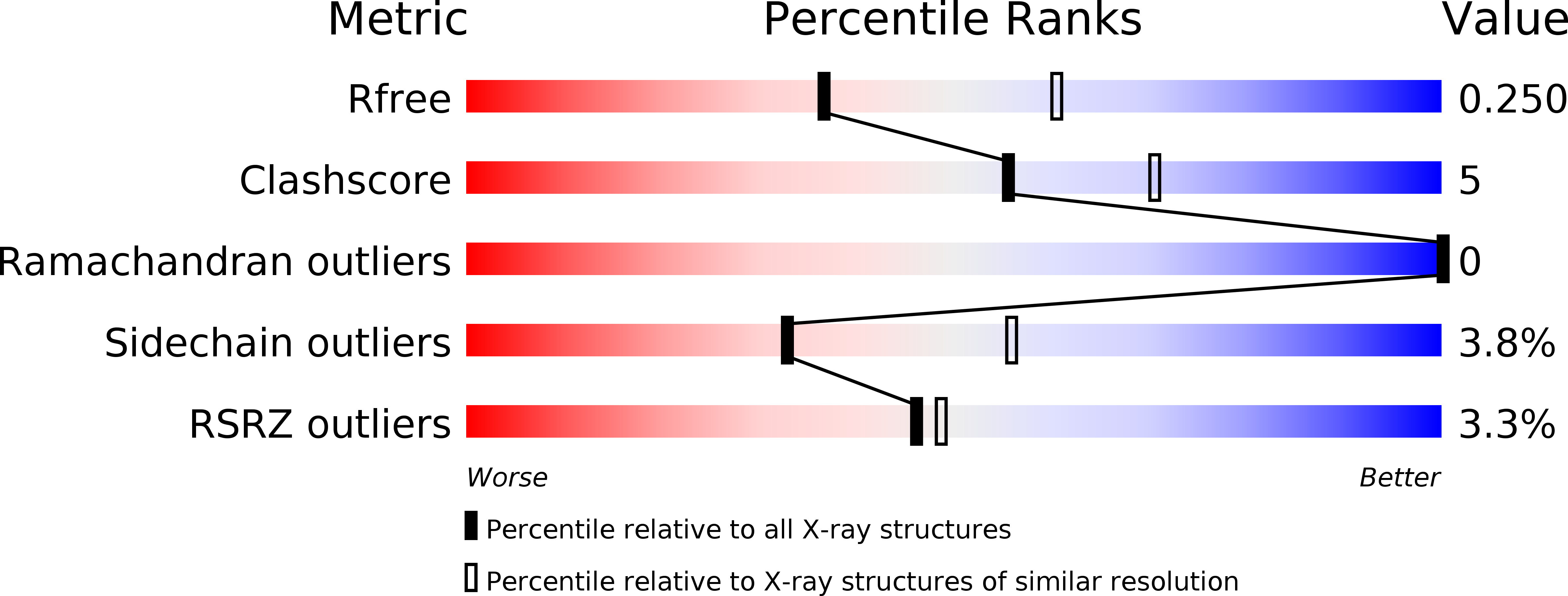
X-ray structure of the ligand-binding core of GluA2 in complex with (S)-7-HPCA at 2.5 A resolution
Biological Source:
Source Organism:
Host Organism:
Method Details:
Experimental Method:
Resolution:
2.48 Å
R-Value Free:
0.26
R-Value Work:
0.17
R-Value Observed:
0.17
Space Group:
P 21 21 2
Macromolecular Entities
Structures with similar UniProt ID
Protein Blast
Polymer Type:polypeptide(L)
Molecule:Glutamate receptor 2
Gene (Uniprot):Gria2
Chain IDs:A, B, C
Chain Length:261
Number of Molecules:3
Biological Source:Rattus norvegicus
Primary Citation
Biostructural and pharmacological studies of bicyclic analogues of the 3-isoxazolol glutamate receptor agonist ibotenic acid.
J. Med. Chem. 53 8354 8361 (2010)
PMID: 21067182 DOI: 10.1021/jm101218a

Abstact

We describe an improved synthesis and detailed pharmacological characterization of the conformationally restricted analogue of the naturally occurring nonselective glutamate receptor agonist ibotenic acid (RS)-3-hydroxy-4,5,6,7-tetrahydroisoxazolo[5,4-c]pyridine-7-carboxylic acid (7-HPCA, 5) at AMPA receptor subtypes. Compound 5 was shown to be a subtype-discriminating agonist at AMPA receptors with higher binding affinity and functional potency at GluA1/2 compared to GluA3/4, unlike the isomeric analogue (RS)-3-hydroxy-4,5,6,7-tetrahydroisoxazolo[5,4-c]pyridine-5-carboxylic acid (5-HPCA, 4) that binds to all AMPA receptor subtypes with comparable potency. Biostructural X-ray crystallographic studies of 4 and 5 reveal different binding modes of (R)-4 and (S)-5 in the GluA2 agonist binding domain. WaterMap analysis of the GluA2 and GluA4 binding pockets with (R)-4 and (S)-5 suggests that the energy of hydration sites is ligand dependent, which may explain the observed selectivity.

Legend

Protein

Chemical

Disease

Primary Citation of related structures
Feedback Form
Name
Email
Institute
Feedback