3PBP image
Deposition Date 2010-10-20
Release Date 2011-10-26
Last Version Date 2024-11-27
Entry Detail
PDB ID:
3PBP
Title:
Structure of the yeast heterotrimeric Nup82-Nup159-Nup116 nucleoporin complex
Biological Source:
Source Organism(s):
Expression System(s):
Method Details:
Experimental Method:
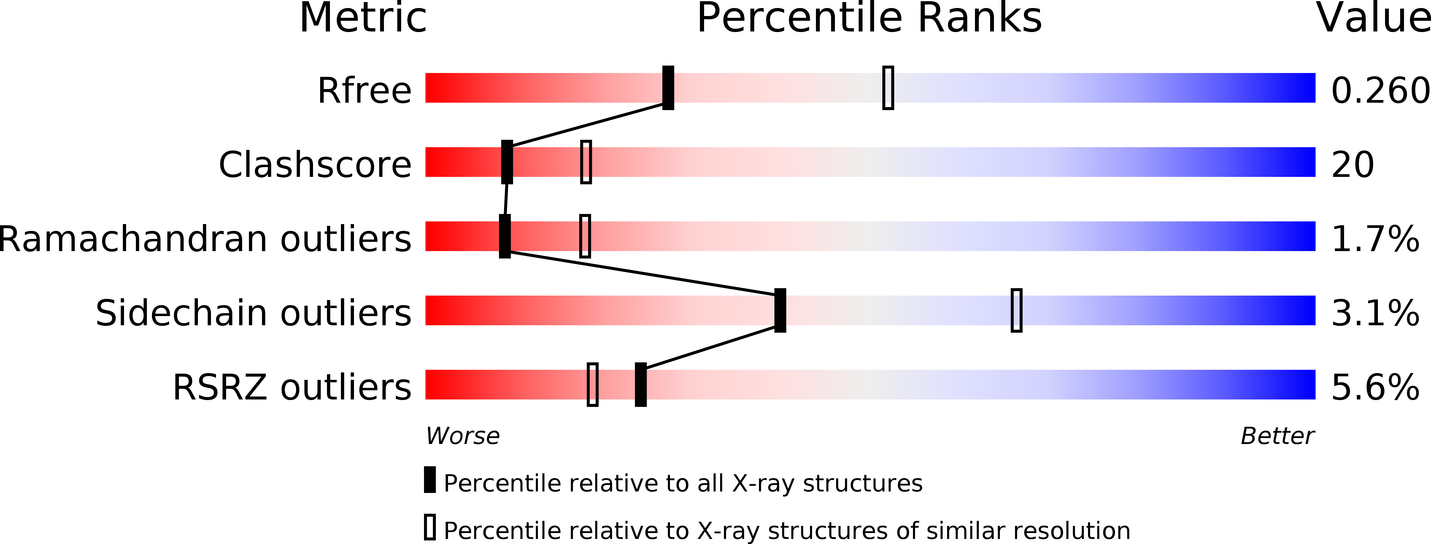
Resolution:
2.60 Å
R-Value Free:
0.27
R-Value Work:
0.25
Space Group:
P 1
Macromolecular Entities
Structures with similar UniProt ID
Protein Blast
Polymer Type:polypeptide(L)
Molecule:Nucleoporin NUP82
Gene (Uniprot):NUP82
Mutagens:C396S
Chain IDs:A, D, G, J
Chain Length:452
Number of Molecules:4
Biological Source:Saccharomyces cerevisiae
Structures with similar UniProt ID
Protein Blast
Polymer Type:polypeptide(L)
Molecule:Nucleoporin NUP116/NSP116
Gene (Uniprot):NUP116
Chain IDs:B, E, H, K
Chain Length:148
Number of Molecules:4
Biological Source:Saccharomyces cerevisiae
Structures with similar UniProt ID
Protein Blast
Polymer Type:polypeptide(L)
Molecule:Nucleoporin NUP159
Gene (Uniprot):NUP159
Chain IDs:C, F, I, L
Chain Length:36
Number of Molecules:4
Biological Source:Saccharomyces cerevisiae
Modified Residue
Compound ID Chain ID Parent Comp ID Details 2D Image
MSE A MET SELENOMETHIONINE
Ligand Molecules
Primary Citation
Structural and functional analysis of an essential nucleoporin heterotrimer on the cytoplasmic face of the nuclear pore complex.
Proc.Natl.Acad.Sci.USA 108 16571 16576 (2011)
PMID: 21930948 DOI: 10.1073/pnas.1112846108

Abstact

So far, only a few of the interactions between the ≈ 30 nucleoporins comprising the modular structure of the nuclear pore complex have been defined at atomic resolution. Here we report the crystal structure, at 2.6 Å resolution, of a heterotrimeric complex, composed of fragments of three cytoplasmically oriented nucleoporins of yeast: Nup82, Nup116, and Nup159. Our data show that the Nup82 fragment, representing more than the N-terminal half of the molecule, folds into an extensively decorated, seven-bladed β-propeller that forms the centerpiece of this heterotrimeric complex and anchors both a C-terminal fragment of Nup116 and the C-terminal tail of Nup159. Binding between Nup116 and Nup82 is mutually reinforced via two loops, one emanating from the Nup82 β-propeller and the other one from the β-sandwich fold of Nup116, each contacting binding pockets in their counterparts. The Nup82-Nup159 interaction occurs through an amphipathic α-helix of Nup159, which is cradled in a large hydrophobic groove that is generated from several large surface decorations of the Nup82 β-propeller. Although Nup159 and Nup116 fragments bind to the Nup82 β-propeller in close vicinity, there are no direct contacts between them, consistent with the noncooperative binding that was detected biochemically. Extensive mutagenesis delineated hot-spot residues for these interactions. We also showed that the Nup82 β-propeller binds to other yeast Nup116 family members, Nup145N, Nup100 and to the mammalian homolog, Nup98. Notably, each of the three nucleoporins contains additional nuclear pore complex binding sites, distinct from those that were defined here in the heterotrimeric Nup82•Nup159•Nup116 complex.

Legend

Protein

Chemical

Disease

Primary Citation of related structures
Feedback Form
Name
Email
Institute
Feedback