3PBL image
Deposition Date 2010-10-20
Release Date 2010-11-03
Last Version Date 2024-10-16
Entry Detail
PDB ID:
3PBL
Title:
Structure of the human dopamine D3 receptor in complex with eticlopride
Biological Source:
Source Organism:
Host Organism:
Method Details:
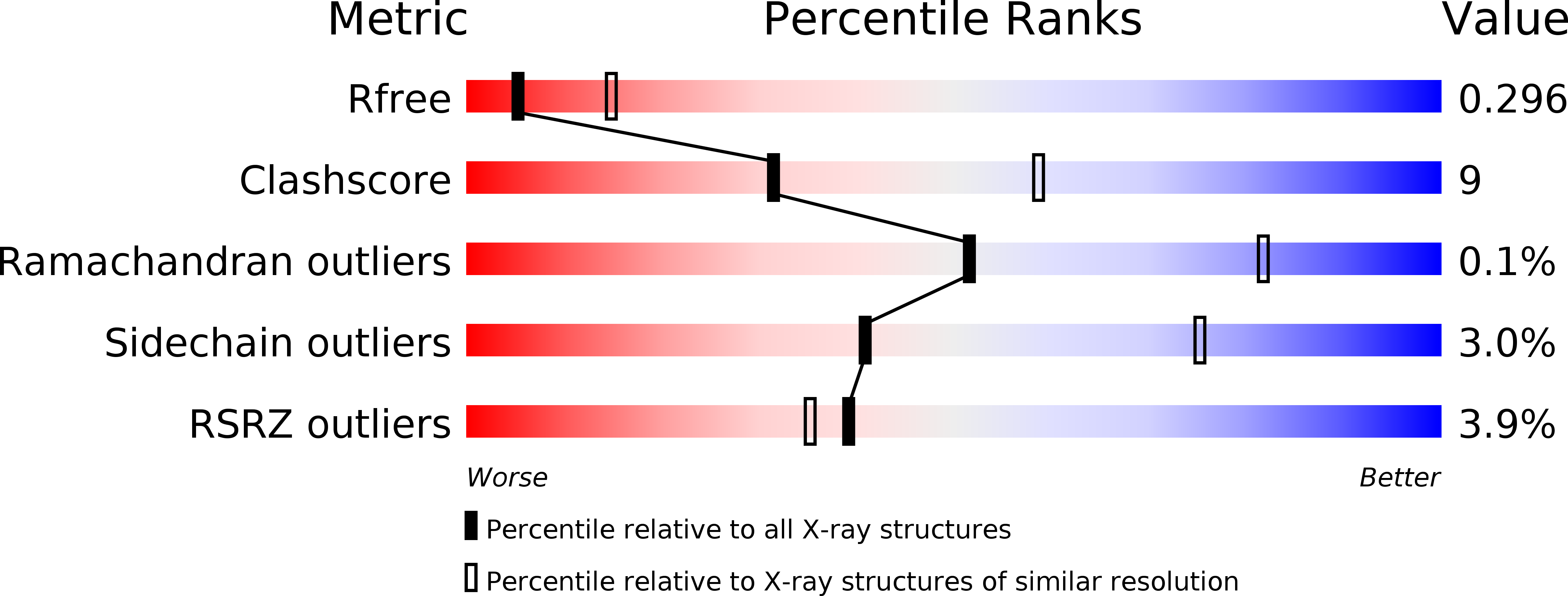
Experimental Method:
Resolution:
2.89 Å
R-Value Free:
0.27
R-Value Work:
0.24
R-Value Observed:
0.24
Space Group:
P 21 21 21
Macromolecular Entities
Polymer Type:polypeptide(L)
Molecule:D(3) dopamine receptor, Lysozyme chimera
Gene (Uniprot):E, DRD3
Mutations:L119W, C1054T, C1097A
Chain IDs:A, B
Chain Length:481
Number of Molecules:2
Biological Source:Homo sapiens, Enterobacteria phage T4
Ligand Molecules
Peptide-like Molecules
PRD_900001
Primary Citation
Structure of the human dopamine d3 receptor in complex with a d2/d3 selective antagonist.
Science 330 1091 1095 (2010)
PMID: 21097933 DOI: 10.1126/science.1197410

Abstact

Dopamine modulates movement, cognition, and emotion through activation of dopamine G protein-coupled receptors in the brain. The crystal structure of the human dopamine D3 receptor (D3R) in complex with the small molecule D2R/D3R-specific antagonist eticlopride reveals important features of the ligand binding pocket and extracellular loops. On the intracellular side of the receptor, a locked conformation of the ionic lock and two distinctly different conformations of intracellular loop 2 are observed. Docking of R-22, a D3R-selective antagonist, reveals an extracellular extension of the eticlopride binding site that comprises a second binding pocket for the aryl amide of R-22, which differs between the highly homologous D2R and D3R. This difference provides direction to the design of D3R-selective agents for treating drug abuse and other neuropsychiatric indications.

Legend

Protein

Chemical

Disease

Primary Citation of related structures