3PBK image
Deposition Date 2010-10-20
Release Date 2010-12-22
Last Version Date 2024-11-27
Entry Detail
PDB ID:
3PBK
Keywords:
Title:
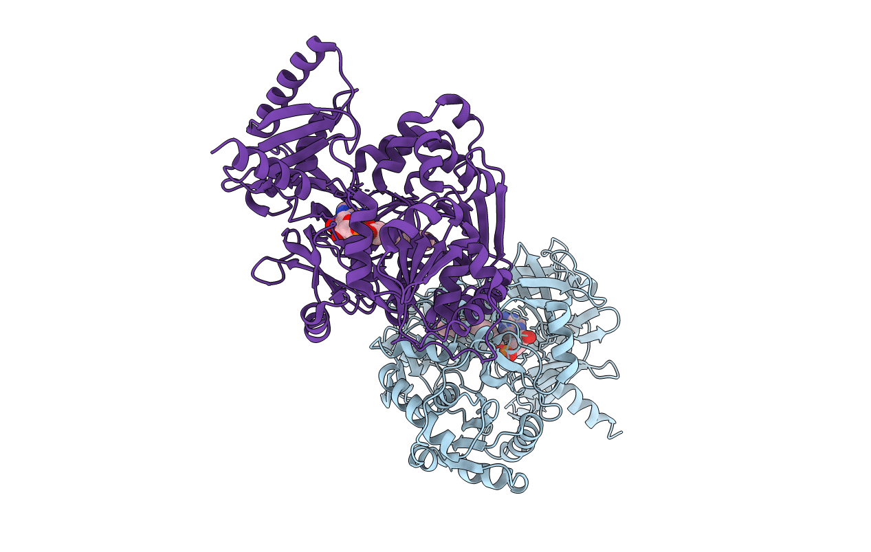
Structural and Functional Studies of Fatty Acyl-Adenylate Ligases from E. coli and L. pneumophila
Biological Source:
Source Organism:
Host Organism:
Method Details:
Experimental Method:
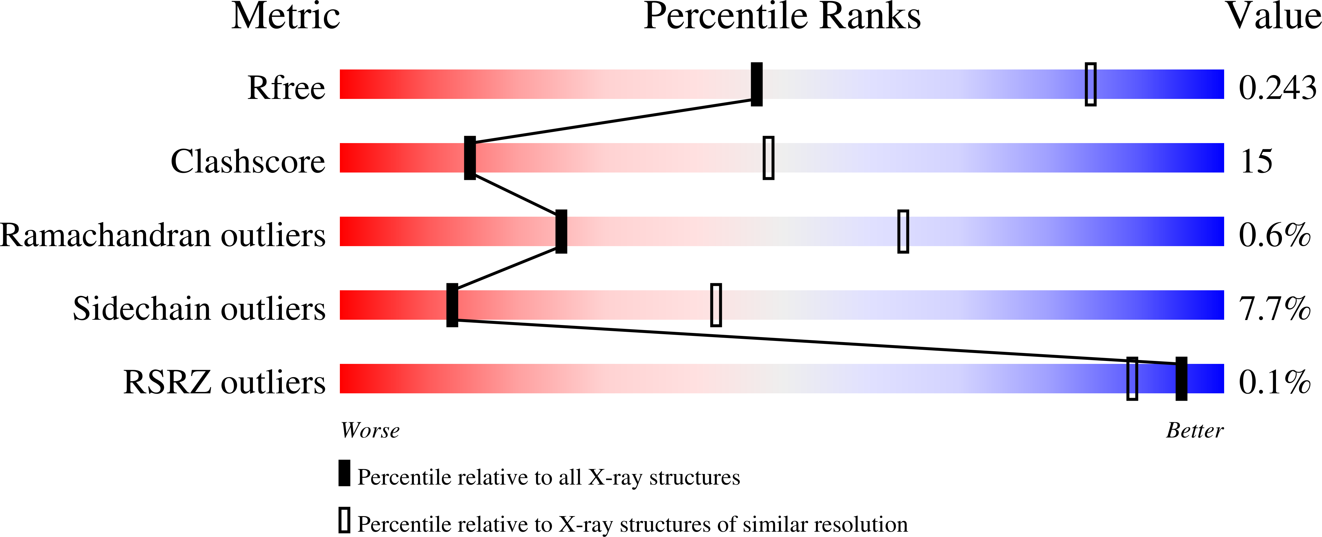
Resolution:
3.00 Å
R-Value Free:
0.25
R-Value Work:
0.19
R-Value Observed:
0.19
Space Group:
P 21 21 21
Macromolecular Entities
Polymer Type:polypeptide(L)
Molecule:Fatty Acyl-Adenylate Ligase
Chain IDs:A, B
Chain Length:583
Number of Molecules:2
Biological Source:Escherichia coli
Modified Residue
Compound ID Chain ID Parent Comp ID Details 2D Image
MSE A MET SELENOMETHIONINE
Ligand Molecules
Primary Citation
Structural and Functional Studies of Fatty Acyl Adenylate Ligases from E. coli and L. pneumophila.
J.Mol.Biol. 406 313 324 (2011)
PMID: 21185305 DOI: 10.1016/j.jmb.2010.12.011

Abstact

Fatty acyl-AMP ligase (FAAL) is a new member of a family of adenylate-forming enzymes that were recently discovered in Mycobacterium tuberculosis. They are similar in sequence to fatty acyl-coenzyme A (CoA) ligases (FACLs). However, while FACLs perform a two-step catalytic reaction, AMP ligation followed by CoA ligation using ATP and CoA as cofactors, FAALs produce only the acyl adenylate and are unable to perform the second step. We report X-ray crystal structures of full-length FAAL from Escherichia coli (EcFAAL) and FAAL from Legionella pneumophila (LpFAAL) bound to acyl adenylate, determined at resolution limits of 3.0 and 1.85 Å, respectively. The structures share a larger N-terminal domain and a smaller C-terminal domain, which together resemble the previously determined structures of FAAL and FACL proteins. Our two structures occur in quite different conformations. EcFAAL adopts the adenylate-forming conformation typical of FACLs, whereas LpFAAL exhibits a unique intermediate conformation. Both EcFAAL and LpFAAL have insertion motifs that distinguish them from the FACLs. Structures of EcFAAL and LpFAAL reveal detailed interactions between this insertion motif and the interdomain hinge region and with the C-terminal domain. We suggest that the insertion motifs support sufficient interdomain motions to allow substrate binding and product release during acyl adenylate formation, but they preclude CoA binding, thereby preventing CoA ligation.

Legend

Protein

Chemical

Disease

Primary Citation of related structures