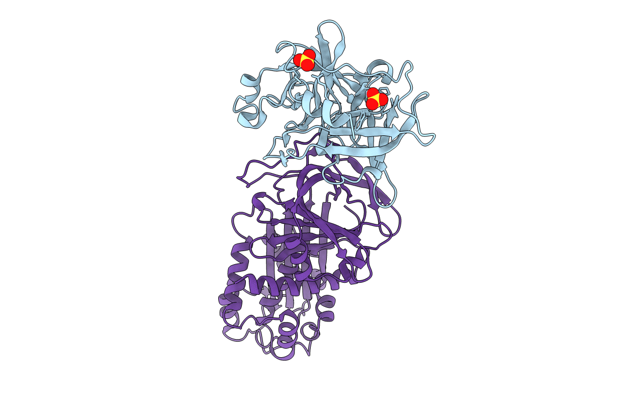
3PB1 image
Entry Detail
PDB ID:
3PB1
Title:
Crystal Structure of a Michaelis Complex between Plasminogen Activator Inhibitor-1 and Urokinase-type Plasminogen Activator
Biological Source:
Source Organism:
Host Organism:
PDB Version:
Deposition Date:
2010-10-20
Release Date:
2010-12-29
Method Details:
Experimental Method:
Resolution:
2.30 Å
R-Value Free:
0.27
R-Value Work:
0.22
R-Value Observed:
0.22
Space Group:
P 32 2 1
Macromolecular Entities
Polymer Type:polypeptide(L)
Description:Plasminogen activator, urokinase
Mutations:S195A
Chain IDs:B (auth: E)
Chain Length:253
Number of Molecules:1
Biological Source:Homo sapiens
Polymer Type:polypeptide(L)
Description:Plasminogen activator inhibitor 1
Mutations:N150H, K154T, Q319L, M354I
Chain IDs:A (auth: I)
Chain Length:379
Number of Molecules:1
Biological Source:Homo sapiens
Ligand Molecules
Primary Citation
Structural basis for recognition of urokinase-type plasminogen activator by plasminogen activator inhibitor-1.
J.Biol.Chem. 286 7027 7032 (2011)
PMID: 21199867 DOI: 10.1074/jbc.M110.204537

Abstact

Plasminogen activator inhibitor-1 (PAI-1), together with its physiological target urokinase-type plasminogen activator (uPA), plays a pivotal role in fibrinolysis, cell migration, and tissue remodeling and is currently recognized as being among the most extensively validated biological prognostic factors in several cancer types. PAI-1 specifically and rapidly inhibits uPA and tissue-type PA (tPA). Despite extensive structural/functional studies on these two reactions, the underlying structural mechanism has remained unknown due to the technical difficulties of obtaining the relevant structures. Here, we report a strategy to generate a PAI-1·uPA(S195A) Michaelis complex and present its crystal structure at 2.3-Å resolution. In this structure, the PAI-1 reactive center loop serves as a bait to attract uPA onto the top of the PAI-1 molecule. The P4-P3' residues of the reactive center loop interact extensively with the uPA catalytic site, accounting for about two-thirds of the total contact area. Besides the active site, almost all uPA exosite loops, including the 37-, 60-, 97-, 147-, and 217-loops, are involved in the interaction with PAI-1. The uPA 37-loop makes an extensive interaction with PAI-1 β-sheet B, and the 147-loop directly contacts PAI-1 β-sheet C. Both loops are important for initial Michaelis complex formation. This study lays down a foundation for understanding the specificity of PAI-1 for uPA and tPA and provides a structural basis for further functional studies.

Legend

Protein

Chemical

Disease

Primary Citation of related structures