3PAH image
Deposition Date 1998-08-20
Release Date 1999-04-27
Last Version Date 2024-11-06
Entry Detail
PDB ID:
3PAH
Keywords:
Title:
HUMAN PHENYLALANINE HYDROXYLASE CATALYTIC DOMAIN DIMER WITH BOUND ADRENALINE INHIBITOR
Biological Source:
Source Organism:
Homo sapiens (Taxon ID: 9606)
Host Organism:
Method Details:
Experimental Method:
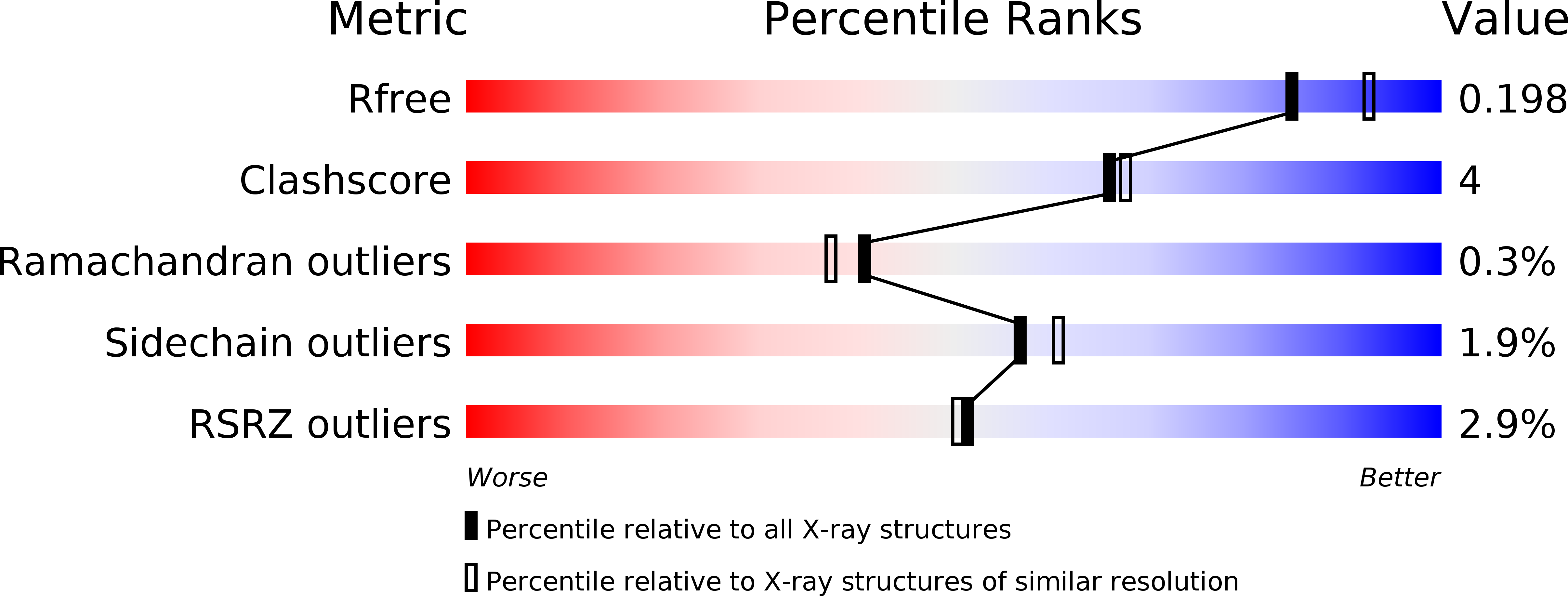
Resolution:
2.00 Å
R-Value Free:
0.20
R-Value Work:
0.17
R-Value Observed:
0.17
Space Group:
C 2 2 21
Macromolecular Entities
Structures with similar UniProt ID
Protein Blast
Polymer Type:polypeptide(L)
Molecule:PHENYLALANINE HYDROXYLASE
Gene (Uniprot):PAH
Chain IDs:A
Chain Length:308
Number of Molecules:1
Biological Source:Homo sapiens
Primary Citation
Crystallographic analysis of the human phenylalanine hydroxylase catalytic domain with bound catechol inhibitors at 2.0 A resolution.
Biochemistry 37 15638 15646 (1998)
PMID: 9843368 DOI: 10.1021/bi9815290

Abstact

The aromatic amino acid hydroxylases represent a superfamily of structurally and functionally closely related enzymes, one of those functions being reversible inhibition by catechol derivatives. Here we present the crystal structure of the dimeric catalytic domain (residues 117-424) of human phenylalanine hydroxylase (hPheOH), cocrystallized with various potent and well-known catechol inhibitors and refined at a resolution of 2.0 A. The catechols bind by bidentate coordination to each iron in both subunits of the dimer through the catechol hydroxyl groups, forming a blue-green colored ligand-to-metal charge-transfer complex. In addition, Glu330 and Tyr325 are identified as determinant residues in the recognition of the inhibitors. In particular, the interaction with Glu330 conforms to the structural explanation for the pH dependence of catecholamine binding to PheOH, with a pKa value of 5.1 (20 degreesC). The overall structure of the catechol-bound enzyme is very similar to that of the uncomplexed enzyme (rms difference of 0.2 A for the Calpha atoms). Most striking is the replacement of two iron-bound water molecules with catechol hydroxyl groups. This change is consistent with a change in the ligand field symmetry of the high-spin (S = 5/2) Fe(III) from a rhombic to a nearly axial ligand field symmetry as seen upon noradrenaline binding using EPR spectroscopy [Martinez, A., Andersson, K. K., Haavik, J., and Flatmark, T. (1991) Eur. J. Biochem. 198, 675-682]. Crystallographic comparison with the structurally related rat tyrosine hydroxylase binary complex with the oxidized cofactor 7,8-dihydrobiopterin revealed overlapping binding sites for the catechols and the cofactor, compatible with a competitive type of inhibition of the catechols versus BH4. The comparison demonstrates some structural differences at the active site as the potential basis for the different substrate specificity of the two enzymes.

Legend

Protein

Chemical

Disease

Primary Citation of related structures