3P7N image
Deposition Date 2010-10-12
Release Date 2011-05-25
Last Version Date 2023-09-06
Entry Detail
PDB ID:
3P7N
Title:
Crystal structure of light activated transcription factor El222 from Erythrobacter litoralis
Biological Source:
Source Organism:
Host Organism:
Method Details:
Experimental Method:
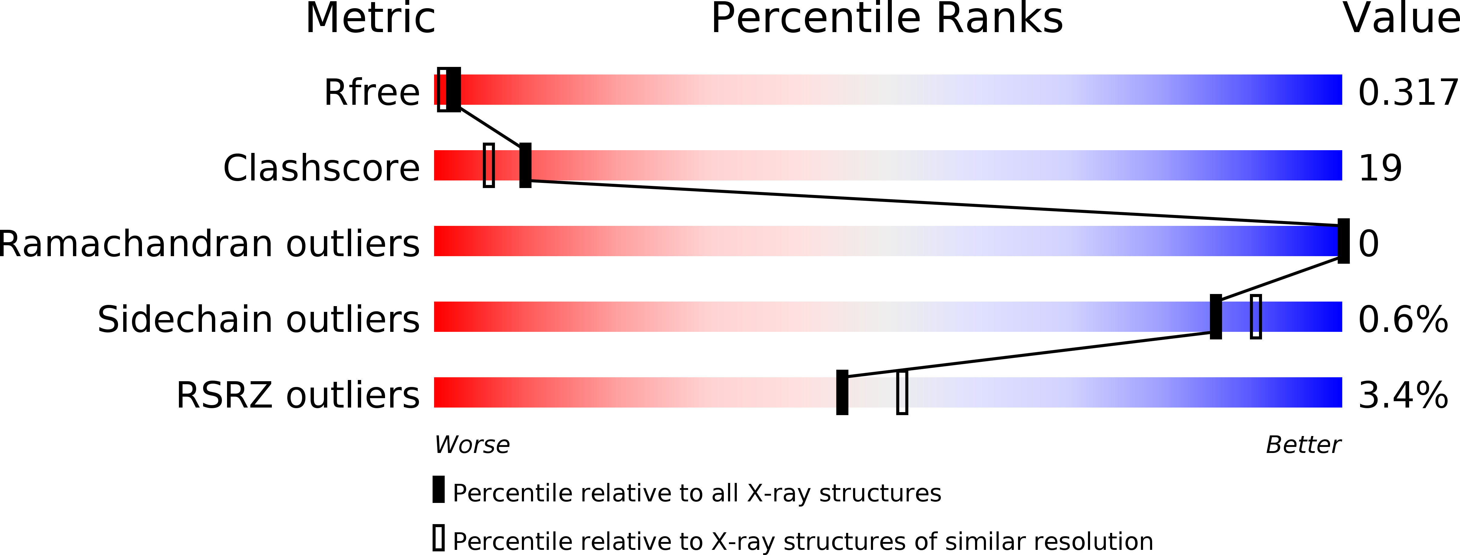
Resolution:
2.10 Å
R-Value Free:
0.32
R-Value Work:
0.26
R-Value Observed:
0.26
Space Group:
P 1 21 1
Macromolecular Entities
Polymer Type:polypeptide(L)
Molecule:Sensor histidine kinase
Gene (Uniprot):ELI_04755
Chain IDs:A, B
Chain Length:258
Number of Molecules:2
Biological Source:Erythrobacter litoralis
Ligand Molecules
Primary Citation
Structural basis of photosensitivity in a bacterial light-oxygen-voltage/helix-turn-helix (LOV-HTH) DNA-binding protein.
Proc.Natl.Acad.Sci.USA 108 9449 9454 (2011)
PMID: 21606338 DOI: 10.1073/pnas.1100262108

Abstact

Light-oxygen-voltage (LOV) domains are blue light-activated signaling modules integral to a wide range of photosensory proteins. Upon illumination, LOV domains form internal protein-flavin adducts that generate conformational changes which control effector function. Here we advance our understanding of LOV regulation with structural, biophysical, and biochemical studies of EL222, a light-regulated DNA-binding protein. The dark-state crystal structure reveals interactions between the EL222 LOV and helix-turn-helix domains that we show inhibit DNA binding. Solution biophysical data indicate that illumination breaks these interactions, freeing the LOV and helix-turn-helix domains of each other. This conformational change has a key functional effect, allowing EL222 to bind DNA in a light-dependent manner. Our data reveal a conserved signaling mechanism among diverse LOV-containing proteins, where light-induced conformational changes trigger activation via a conserved interaction surface.

Legend

Protein

Chemical

Disease

Primary Citation of related structures