3P63 image
Deposition Date 2010-10-11
Release Date 2011-02-09
Last Version Date 2023-11-01
Entry Detail
PDB ID:
3P63
Title:
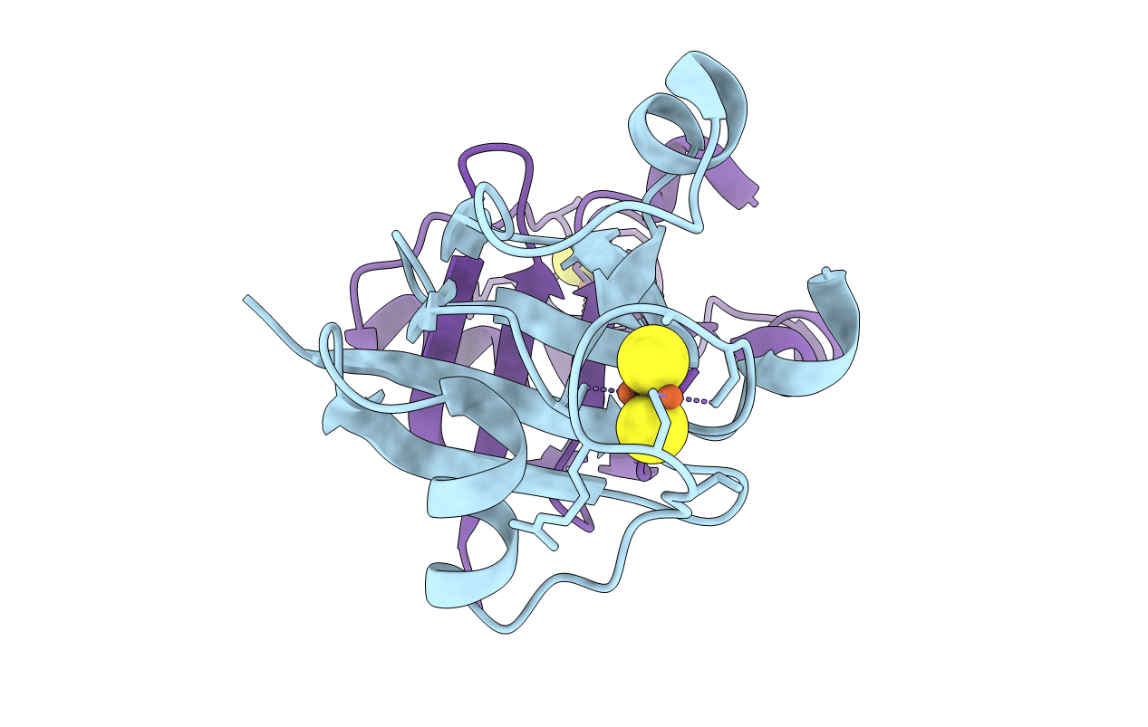
Structure of M. laminosus Ferredoxin with a shorter L1,2 loop
Biological Source:
Source Organism(s):
Expression System(s):
Method Details:
Experimental Method:
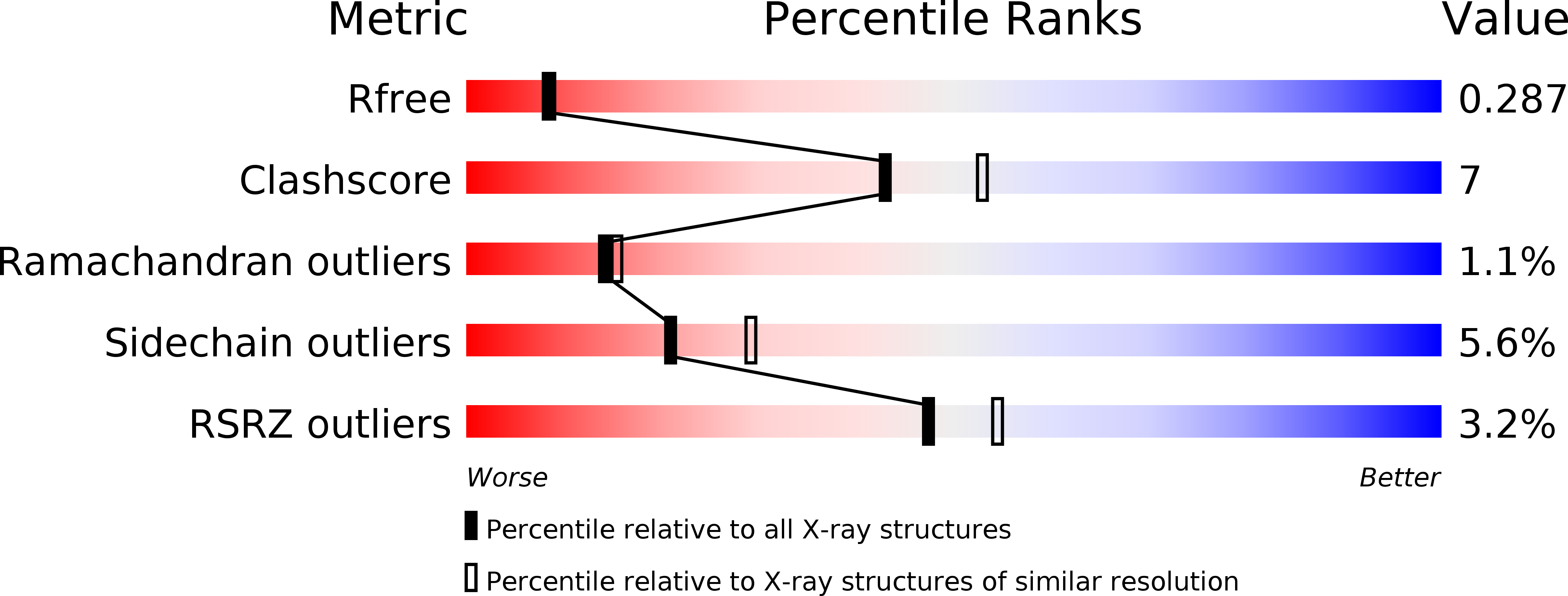
Resolution:
2.30 Å
R-Value Free:
0.28
R-Value Work:
0.21
R-Value Observed:
0.22
Space Group:
P 41
Macromolecular Entities
Structures with similar UniProt ID
Protein Blast
Polymer Type:polypeptide(L)
Molecule:Ferredoxin
Gene (Uniprot):petF
Chain IDs:A, B
Chain Length:96
Number of Molecules:2
Biological Source:Mastigocladus laminosus
Ligand Molecules
Primary Citation
Allostery in the ferredoxin protein motif does not involve a conformational switch.
Proc.Natl.Acad.Sci.USA 108 2240 2245 (2011)
PMID: 21266547 DOI: 10.1073/pnas.1019502108

Abstact

Regulation of protein function via cracking, or local unfolding and refolding of substructures, is becoming a widely recognized mechanism of functional control. Oftentimes, cracking events are localized to secondary and tertiary structure interactions between domains that control the optimal position for catalysis and/or the formation of protein complexes. Small changes in free energy associated with ligand binding, phosphorylation, etc., can tip the balance and provide a regulatory functional switch. However, understanding the factors controlling function in single-domain proteins is still a significant challenge to structural biologists. We investigated the functional landscape of a single-domain plant-type ferredoxin protein and the effect of a distal loop on the electron-transfer center. We find the global stability and structure are minimally perturbed with mutation, whereas the functional properties are altered. Specifically, truncating the L1,2 loop does not lead to large-scale changes in the structure, determined via X-ray crystallography. Further, the overall thermal stability of the protein is only marginally perturbed by the mutation. However, even though the mutation is distal to the iron-sulfur cluster (∼20 Å), it leads to a significant change in the redox potential of the iron-sulfur cluster (57 mV). Structure-based all-atom simulations indicate correlated dynamical changes between the surface-exposed loop and the iron-sulfur cluster-binding region. Our results suggest intrinsic communication channels within the ferredoxin fold, composed of many short-range interactions, lead to the propagation of long-range signals. Accordingly, protein interface interactions that involve L1,2 could potentially signal functional changes in distal regions, similar to what is observed in other allosteric systems.

Legend

Protein

Chemical

Disease

Primary Citation of related structures
Feedback Form
Name
Email
Institute
Feedback