3P5N image
Deposition Date 2010-10-09
Release Date 2010-11-03
Last Version Date 2024-03-20
Entry Detail
PDB ID:
3P5N
Title:
Structure and mechanism of the S component of a bacterial ECF transporter
Biological Source:
Source Organism:
Host Organism:
Method Details:
Experimental Method:
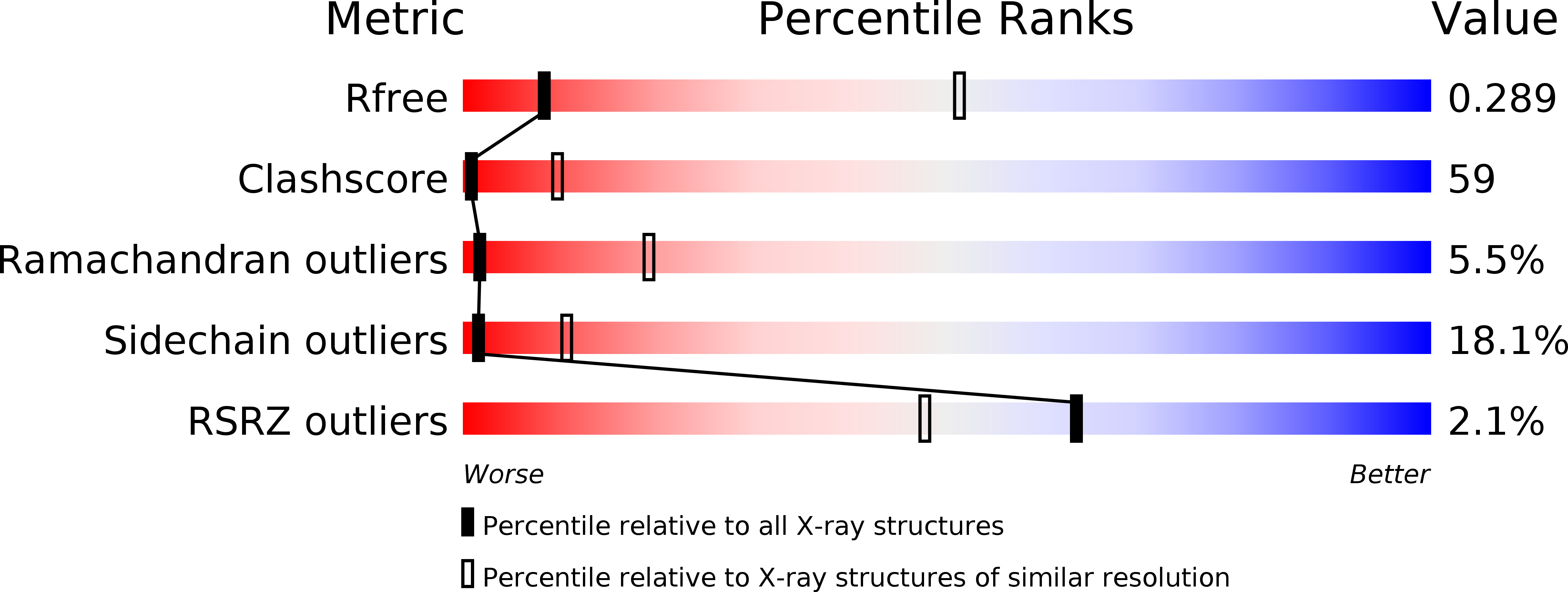
Resolution:
3.60 Å
R-Value Free:
0.28
R-Value Work:
0.26
R-Value Observed:
0.26
Space Group:
P 21 21 21
Macromolecular Entities
Polymer Type:polypeptide(L)
Molecule:Riboflavin uptake protein
Chain IDs:A, B
Chain Length:189
Number of Molecules:2
Biological Source:Staphylococcus aureus
Ligand Molecules
Primary Citation
Structure and mechanism of the S component of a bacterial ECF transporter
Nature 468 717 720 (2010)
PMID: 20972419 DOI: 10.1038/nature09488

Abstact

The energy-coupling factor (ECF) transporters, responsible for vitamin uptake in prokaryotes, are a unique family of membrane transporters. Each ECF transporter contains a membrane-embedded, substrate-binding protein (known as the S component), an energy-coupling module that comprises two ATP-binding proteins (known as the A and A' components) and a transmembrane protein (known as the T component). The structure and transport mechanism of the ECF family remain unknown. Here we report the crystal structure of RibU, the S component of the ECF-type riboflavin transporter from Staphylococcus aureus at 3.6-Å resolution. RibU contains six transmembrane segments, adopts a previously unreported transporter fold and contains a riboflavin molecule bound to the L1 loop and the periplasmic portion of transmembrane segments 4-6. Structural analysis reveals the essential ligand-binding residues, identifies the putative transport path and, with sequence alignment, uncovers conserved structural features and suggests potential mechanisms of action among the ECF transporters.

Legend

Protein

Chemical

Disease

Primary Citation of related structures