3P5D image
Entry Detail
PDB ID:
3P5D
Title:
Structure of the carbohydrate-recognition domain of human Langerin with man5 (Man alpha1-3(Man alpha1-6)Man alpha1-6)(Man- alpha1-3)Man
Biological Source:
Source Organism:
Host Organism:
PDB Version:
Deposition Date:
2010-10-08
Release Date:
2010-12-08
Method Details:
Experimental Method:
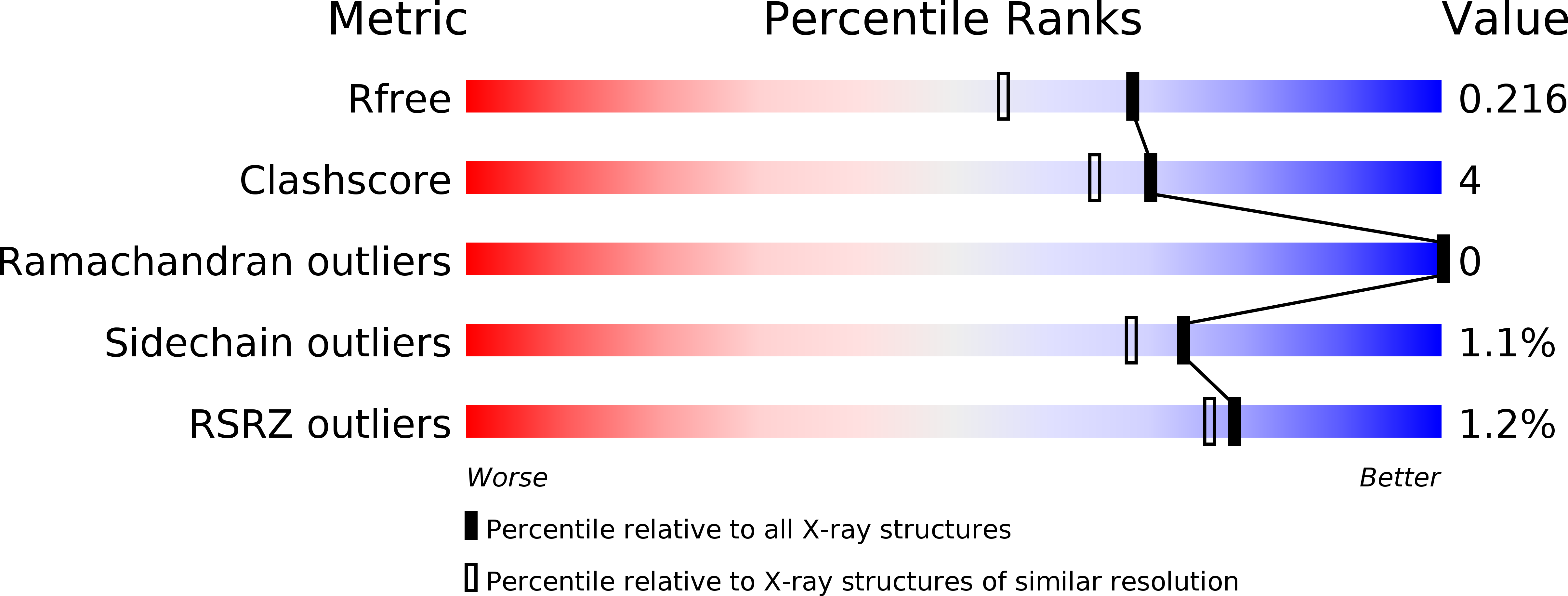
Resolution:
1.80 Å
R-Value Free:
0.22
R-Value Work:
0.17
R-Value Observed:
0.17
Space Group:
P 42
Macromolecular Entities
Polymer Type:polypeptide(L)
Description:C-type lectin domain family 4 member K
Chain IDs:A, B, C, D
Chain Length:136
Number of Molecules:4
Biological Source:Homo sapiens
Peptide-like Molecules
PRD_900112
Primary Citation
Structural Basis for Langerin Recognition of Diverse Pathogen and Mammalian Glycans through a Single Binding Site.
J.Mol.Biol. 405 1027 1039 (2011)
PMID: 21112338 DOI: 10.1016/j.jmb.2010.11.039

Abstact

Langerin mediates the carbohydrate-dependent uptake of pathogens by Langerhans cells in the first step of antigen presentation to the adaptive immune system. Langerin binds to an unusually diverse number of endogenous and pathogenic cell surface carbohydrates, including mannose-containing O-specific polysaccharides derived from bacterial lipopolysaccharides identified here by probing a microarray of bacterial polysaccharides. Crystal structures of the carbohydrate-recognition domain from human langerin bound to a series of oligomannose compounds, the blood group B antigen, and a fragment of β-glucan reveal binding to mannose, fucose, and glucose residues by Ca(2+) coordination of vicinal hydroxyl groups with similar stereochemistry. Oligomannose compounds bind through a single mannose residue, with no other mannose residues contacting the protein directly. There is no evidence for a second Ca(2+)-independent binding site. Likewise, a β-glucan fragment, Glcβ1-3Glcβ1-3Glc, binds to langerin through the interaction of a single glucose residue with the Ca(2+) site. The fucose moiety of the blood group B trisaccharide Galα1-3(Fucα1-2)Gal also binds to the Ca(2+) site, and selective binding to this glycan compared to other fucose-containing oligosaccharides results from additional favorable interactions of the nonreducing terminal galactose, as well as of the fucose residue. Surprisingly, the equatorial 3-OH group and the axial 4-OH group of the galactose residue in 6SO(4)-Galβ1-4GlcNAc also coordinate Ca(2+), a heretofore unobserved mode of galactose binding in a C-type carbohydrate-recognition domain bearing the Glu-Pro-Asn signature motif characteristic of mannose binding sites. Salt bridges between the sulfate group and two lysine residues appear to compensate for the nonoptimal binding of galactose at this site.

Legend

Protein

Chemical

Disease

Primary Citation of related structures