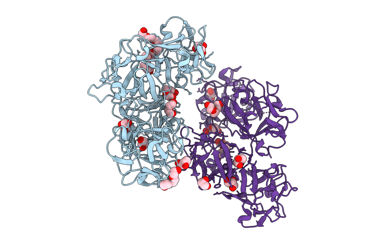
3P53 image
Entry Detail
PDB ID:
3P53
Title:
Structure of fascin
Biological Source:
Source Organism:
Host Organism:
PDB Version:
Deposition Date:
2010-10-07
Release Date:
2011-06-29
Method Details:
Experimental Method:
Resolution:
2.00 Å
R-Value Free:
0.22
R-Value Work:
0.17
R-Value Observed:
0.17
Space Group:
C 1 2 1
Macromolecular Entities
Polymer Type:polypeptide(L)
Description:Fascin
Chain IDs:A, B
Chain Length:496
Number of Molecules:2
Biological Source:Homo sapiens
Primary Citation
Mechanism of actin filament bundling by fascin.
J.Biol.Chem. 286 30087 30096 (2011)
PMID: 21685497 DOI: 10.1074/jbc.M111.251439

Abstact

Fascin is the main actin filament bundling protein in filopodia. Because of the important role filopodia play in cell migration, fascin is emerging as a major target for cancer drug discovery. However, an understanding of the mechanism of bundle formation by fascin is critically lacking. Fascin consists of four β-trefoil domains. Here, we show that fascin contains two major actin-binding sites, coinciding with regions of high sequence conservation in β-trefoil domains 1 and 3. The site in β-trefoil-1 is located near the binding site of the fascin inhibitor macroketone and comprises residue Ser-39, whose phosphorylation by protein kinase C down-regulates actin bundling and formation of filopodia. The site in β-trefoil-3 is related by pseudo-2-fold symmetry to that in β-trefoil-1. The two sites are ∼5 nm apart, resulting in a distance between actin filaments in the bundle of ∼8.1 nm. Residue mutations in both sites disrupt bundle formation in vitro as assessed by co-sedimentation with actin and electron microscopy and severely impair formation of filopodia in cells as determined by rescue experiments in fascin-depleted cells. Mutations of other areas of the fascin surface also affect actin bundling and formation of filopodia albeit to a lesser extent, suggesting that, in addition to the two major actin-binding sites, fascin makes secondary contacts with other filaments in the bundle. In a high resolution crystal structure of fascin, molecules of glycerol and polyethylene glycol are bound in pockets located within the two major actin-binding sites. These molecules could guide the rational design of new anticancer fascin inhibitors.

Legend

Protein

Chemical

Disease

Primary Citation of related structures