3P49 image
Deposition Date 2010-10-06
Release Date 2011-04-06
Last Version Date 2024-02-21
Entry Detail
PDB ID:
3P49
Title:
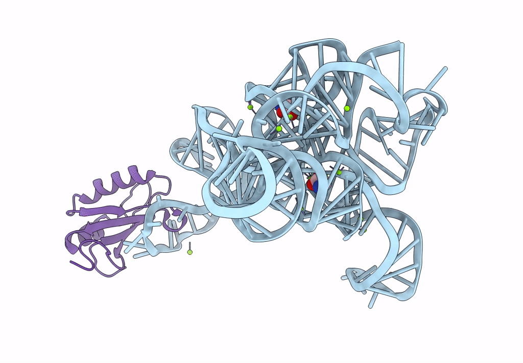
Crystal Structure of a Glycine Riboswitch from Fusobacterium nucleatum
Biological Source:
Source Organism:
Method Details:
Experimental Method:
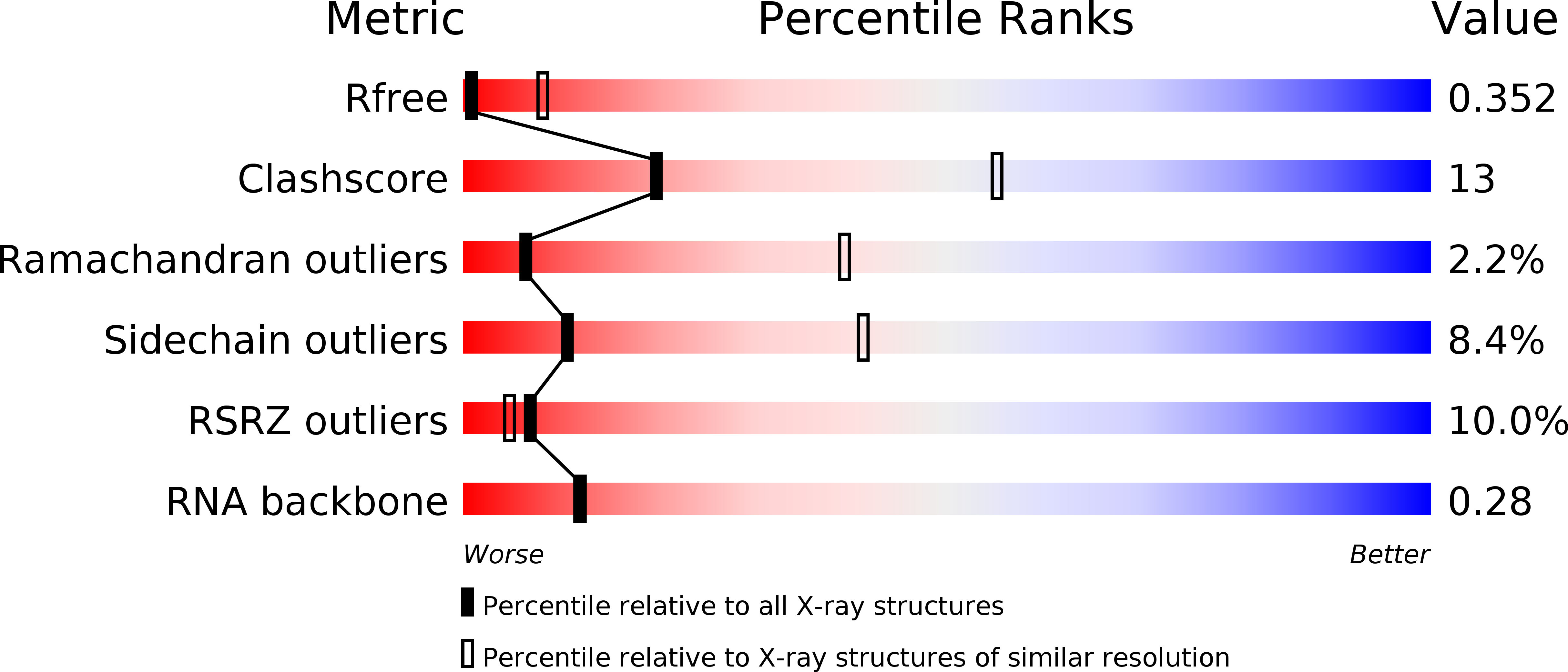
Resolution:
3.55 Å
R-Value Free:
0.30
R-Value Work:
0.28
R-Value Observed:
0.28
Space Group:
P 21 21 21
Macromolecular Entities
Polymer Type:polyribonucleotide
Molecule:GLYCINE RIBOSWITCH
Chain IDs:A
Chain Length:169
Number of Molecules:1
Biological Source:Fusobacterium nucleatum subsp. polymorphum
Structures with similar UniProt ID
Protein Blast
Polymer Type:polypeptide(L)
Molecule:U1 small nuclear ribonucleoprotein A
Gene (Uniprot):SNRPA
Mutagens:Y31H Q36R
Chain IDs:B
Chain Length:98
Number of Molecules:1
Biological Source:Homo sapiens
Primary Citation
Structural basis of cooperative ligand binding by the glycine riboswitch.
Chem.Biol. 18 293 298 (2011)
PMID: 21439473 DOI: 10.1016/j.chembiol.2011.01.013

Abstact

The glycine riboswitch regulates gene expression through the cooperative recognition of its amino acid ligand by a tandem pair of aptamers. A 3.6 Å crystal structure of the tandem riboswitch from the glycine permease operon of Fusobacterium nucleatum reveals the glycine binding sites and an extensive network of interactions, largely mediated by asymmetric A-minor contacts, that serve to communicate ligand binding status between the aptamers. These interactions provide a structural basis for how the glycine riboswitch cooperatively regulates gene expression.

Legend

Protein

Chemical

Disease

Primary Citation of related structures