Deposition Date
2010-09-27
Release Date
2011-02-02
Last Version Date
2025-03-26
Entry Detail
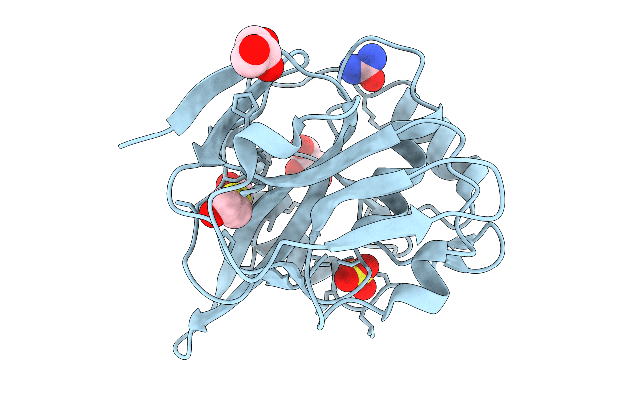
PDB ID:
3P06
Keywords:
Title:
Crystal structure of Tellina virus 1 VP4 protease in the form of an intra-molecular(cis)acyl-enzyme complex.
Biological Source:
Source Organism(s):
Tellina virus 1 (Taxon ID: 321302)
Expression System(s):
Method Details:
Experimental Method:
Resolution:
2.10 Å
R-Value Free:
0.23
R-Value Work:
0.18
R-Value Observed:
0.18
Space Group:
P 64 2 2