
Deposition Date
2010-09-27
Release Date
2011-03-16
Last Version Date
2023-09-06
Entry Detail
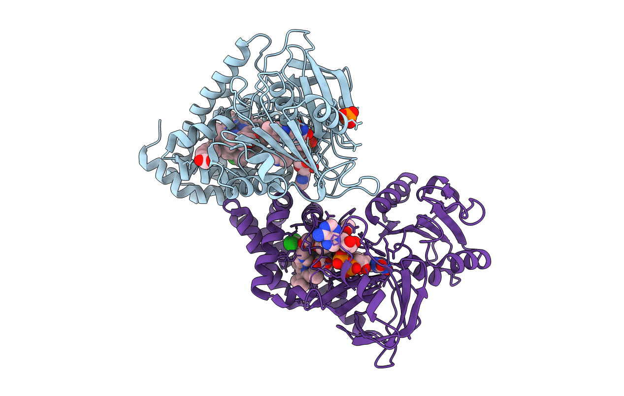
PDB ID:
3OZW
Keywords:
Title:
The Crystal Structure of flavohemoglobin from R. eutrophus in complex with ketoconazole
Biological Source:
Source Organism(s):
Ralstonia eutropha (Taxon ID: 381666)
Expression System(s):
Method Details:
Experimental Method:
Resolution:
2.30 Å
R-Value Free:
0.24
R-Value Work:
0.20
R-Value Observed:
0.20
Space Group:
P 43 21 2
