Deposition Date
2010-09-22
Release Date
2011-02-16
Last Version Date
2024-10-30
Entry Detail
PDB ID:
3OXU
Keywords:
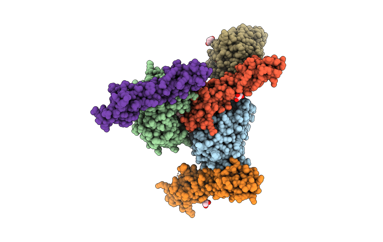
Title:
Complement components factor H CCP19-20 and C3d in complex
Biological Source:
Source Organism(s):
Homo sapiens (Taxon ID: 9606)
Expression System(s):
Method Details:
Experimental Method:
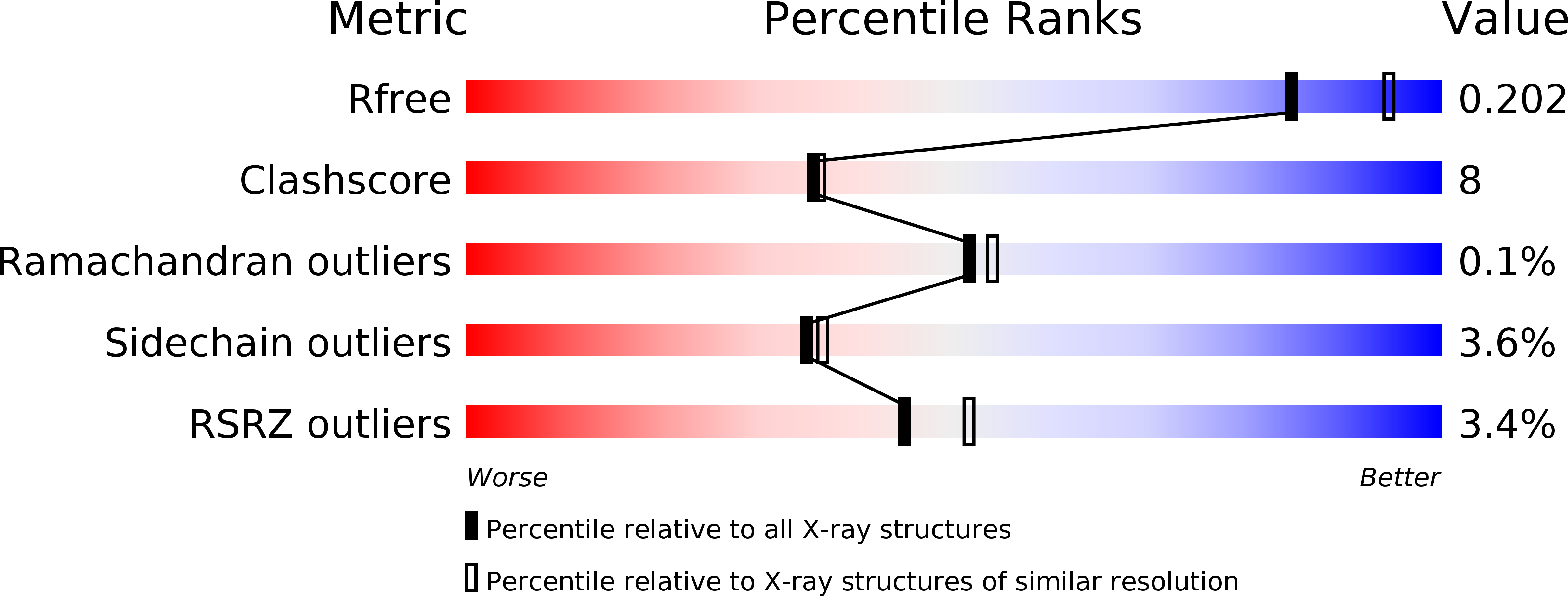
Resolution:
2.10 Å
R-Value Free:
0.20
R-Value Work:
0.17
R-Value Observed:
0.17
Space Group:
P 1