Deposition Date
2010-09-21
Release Date
2010-12-22
Last Version Date
2024-02-21
Entry Detail
PDB ID:
3OX5
Keywords:
Title:
Crystal Structure of the calcium sensor calcium-binding protein 1 (CaBP1)
Biological Source:
Source Organism(s):
Homo sapiens (Taxon ID: 9606)
Expression System(s):
Method Details:
Experimental Method:
Resolution:
2.90 Å
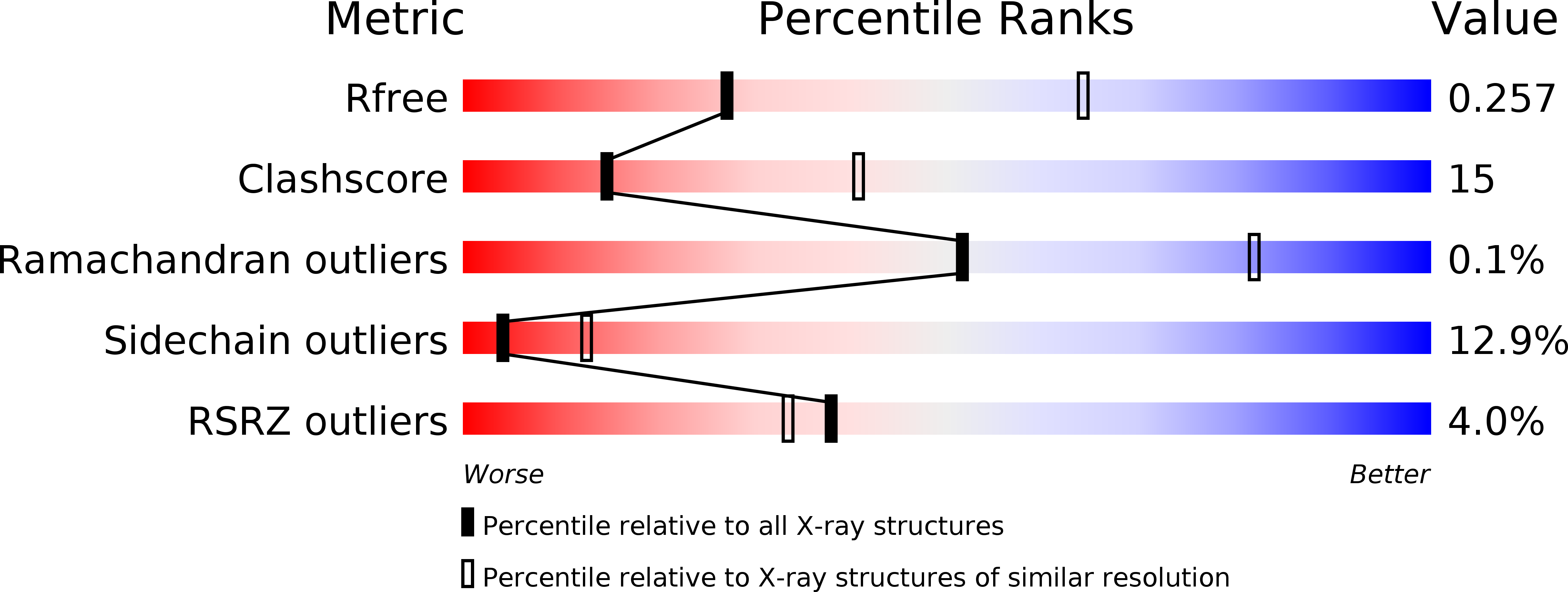
R-Value Free:
0.25
R-Value Work:
0.21
R-Value Observed:
0.21
Space Group:
I 2 3