3OUO image
Deposition Date 2010-09-15
Release Date 2011-05-25
Last Version Date 2024-11-20
Entry Detail
PDB ID:
3OUO
Keywords:
Title:
Structure of the Nucleoprotein from Rift Valley Fever Virus
Biological Source:
Source Organism:
Host Organism:
Method Details:
Experimental Method:
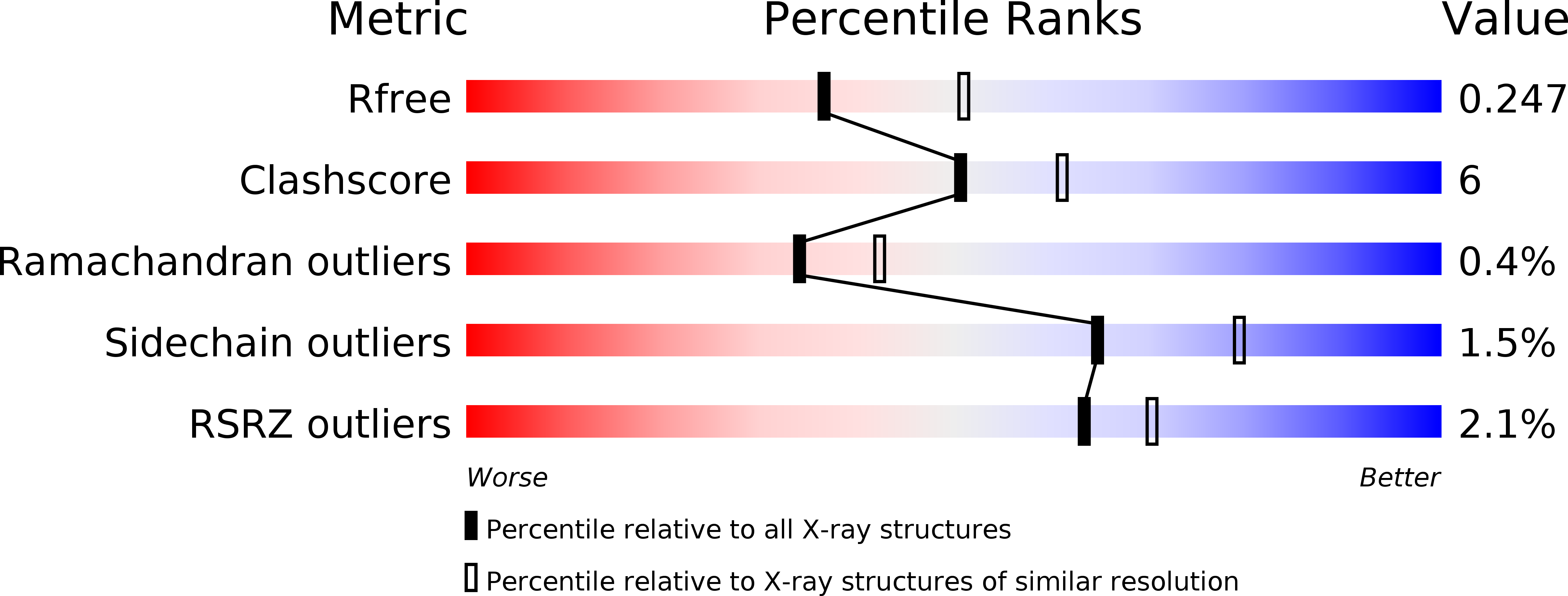
Resolution:
2.30 Å
R-Value Free:
0.24
R-Value Work:
0.19
R-Value Observed:
0.19
Space Group:
P 6
Macromolecular Entities
Polymer Type:polypeptide(L)
Molecule:Nucleoprotein
Gene (Uniprot):N
Chain IDs:A, B, C
Chain Length:245
Number of Molecules:3
Biological Source:Rift valley fever virus
Modified Residue
Compound ID Chain ID Parent Comp ID Details 2D Image
MSE A MET SELENOMETHIONINE
Ligand Molecules
Primary Citation
The hexamer structure of Rift Valley fever virus nucleoprotein suggests a mechanism for its assembly into ribonucleoprotein complexes
Plos Pathog. 7 e1002030 e1002030 (2011)
PMID: 21589902 DOI: 10.1371/journal.ppat.1002030

Abstact

Rift Valley fever virus (RVFV), a Phlebovirus with a genome consisting of three single-stranded RNA segments, is spread by infected mosquitoes and causes large viral outbreaks in Africa. RVFV encodes a nucleoprotein (N) that encapsidates the viral RNA. The N protein is the major component of the ribonucleoprotein complex and is also required for genomic RNA replication and transcription by the viral polymerase. Here we present the 1.6 Å crystal structure of the RVFV N protein in hexameric form. The ring-shaped hexamers form a functional RNA binding site, as assessed by mutagenesis experiments. Electron microscopy (EM) demonstrates that N in complex with RNA also forms rings in solution, and a single-particle EM reconstruction of a hexameric N-RNA complex is consistent with the crystallographic N hexamers. The ring-like organization of the hexamers in the crystal is stabilized by circular interactions of the N terminus of RVFV N, which forms an extended arm that binds to a hydrophobic pocket in the core domain of an adjacent subunit. The conformation of the N-terminal arm differs from that seen in a previous crystal structure of RVFV, in which it was bound to the hydrophobic pocket in its own core domain. The switch from an intra- to an inter-molecular interaction mode of the N-terminal arm may be a general principle that underlies multimerization and RNA encapsidation by N proteins from Bunyaviridae. Furthermore, slight structural adjustments of the N-terminal arm would allow RVFV N to form smaller or larger ring-shaped oligomers and potentially even a multimer with a super-helical subunit arrangement. Thus, the interaction mode between subunits seen in the crystal structure would allow the formation of filamentous ribonucleocapsids in vivo. Both the RNA binding cleft and the multimerization site of the N protein are promising targets for the development of antiviral drugs.

Legend

Protein

Chemical

Disease

Primary Citation of related structures