Deposition Date
2010-09-09
Release Date
2011-08-03
Last Version Date
2023-09-06
Entry Detail
PDB ID:
3OSS
Keywords:
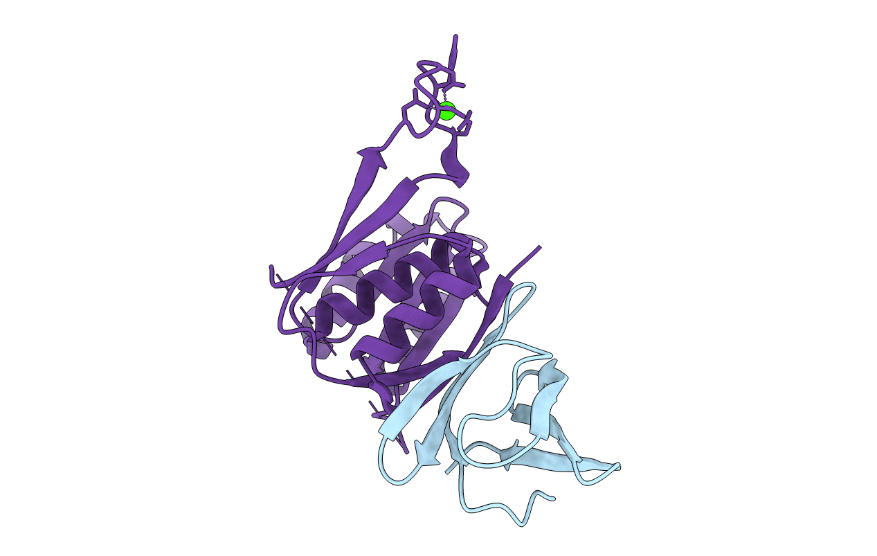
Title:
The crystal structure of enterotoxigenic Escherichia coli GspC-GspD complex from the type II secretion system
Biological Source:
Source Organism(s):
Escherichia coli (Taxon ID: 316401)
Expression System(s):
Method Details:
Experimental Method:
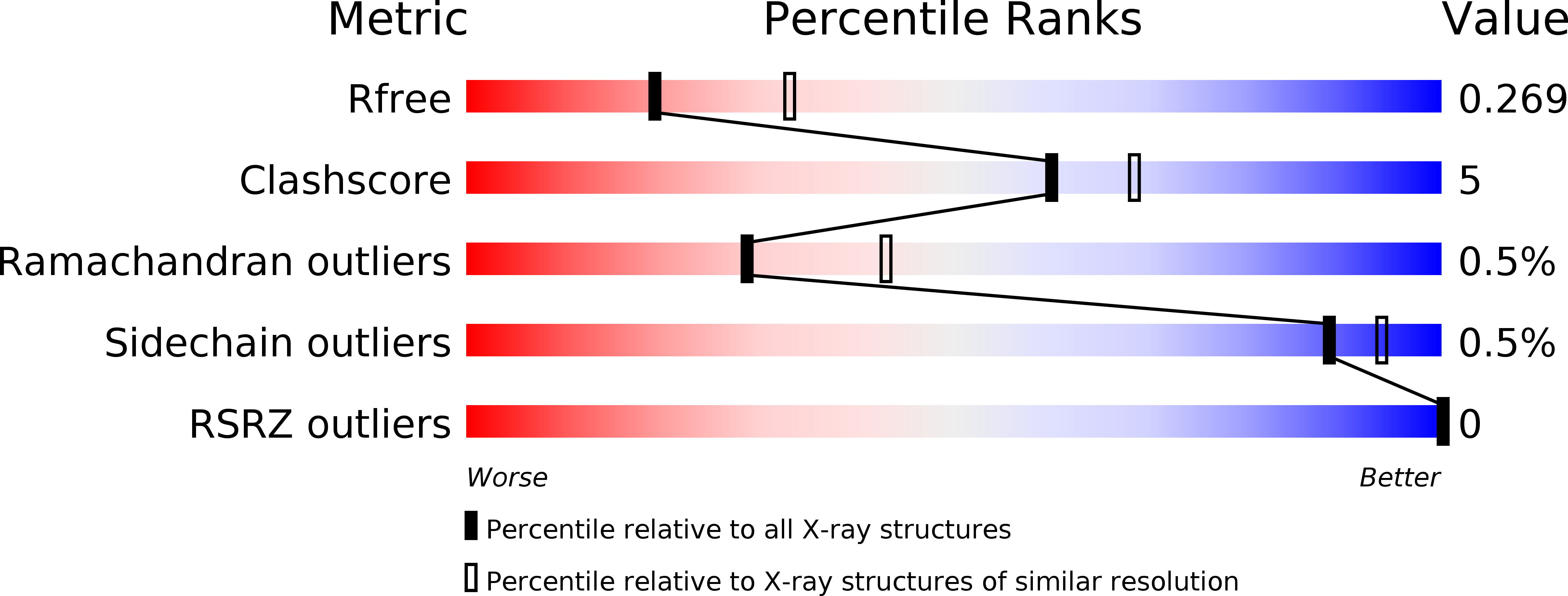
Resolution:
2.63 Å
R-Value Free:
0.26
R-Value Work:
0.21
R-Value Observed:
0.21
Space Group:
P 21 21 21