3ORF image
Deposition Date 2010-09-07
Release Date 2011-07-27
Last Version Date 2023-11-01
Entry Detail
PDB ID:
3ORF
Keywords:
Title:
Crystal Structure of Dihydropteridine Reductase from Dictyostelium discoideum
Biological Source:
Source Organism:
Host Organism:
Method Details:
Experimental Method:
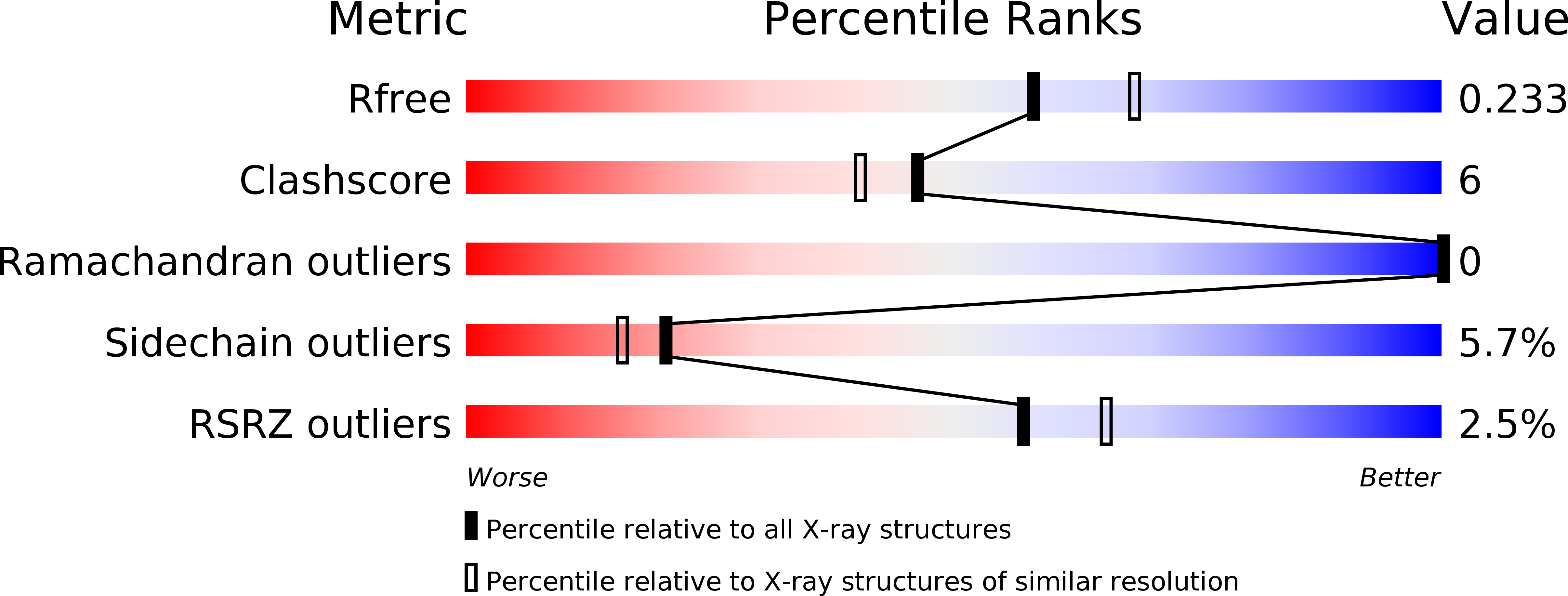
Resolution:
2.16 Å
R-Value Free:
0.24
R-Value Work:
0.18
R-Value Observed:
0.18
Space Group:
P 1 21 1
Macromolecular Entities
Polymer Type:polypeptide(L)
Molecule:Dihydropteridine reductase
Gene (Uniprot):qdpr
Chain IDs:A, B, C, D
Chain Length:251
Number of Molecules:4
Biological Source:Dictyostelium discoideum
Ligand Molecules
Primary Citation
Structural insights into the dual substrate specificities of mammalian and Dictyostelium dihydropteridine reductases toward two stereoisomers of quinonoid dihydrobiopterin
Febs Lett. 585 2640 2646 (2011)
PMID: 21819985 DOI: 10.1016/j.febslet.2011.07.018

Abstact

Up to now, d-threo-tetrahydrobiopterin (DH(4), dictyopterin) was detected only in Dictyostelium discoideum, while the isomer L-erythro-tetrahydrobioterin (BH(4)) is common in mammals. To elucidate the mechanism of DH(4) regeneration by D. discoideum dihydropteridine reductase (DicDHPR), we have determined the crystal structure of DicDHPR complexed with NAD(+) at 2.16 Å resolution. Significant structural differences from mammalian DHPRs are found around the coenzyme binding site, resulting in a higher K(m) value for NADH (K(m)=46.51±0.4 μM) than mammals. In addition, we have found that rat DHPR as well as DicDHPR could bind to both substrates quinonoid-BH(2) and quinonoid-DH(2) by docking calculations and have confirmed their catalytic activity by in vitro assay.

Legend

Protein

Chemical

Disease

Primary Citation of related structures