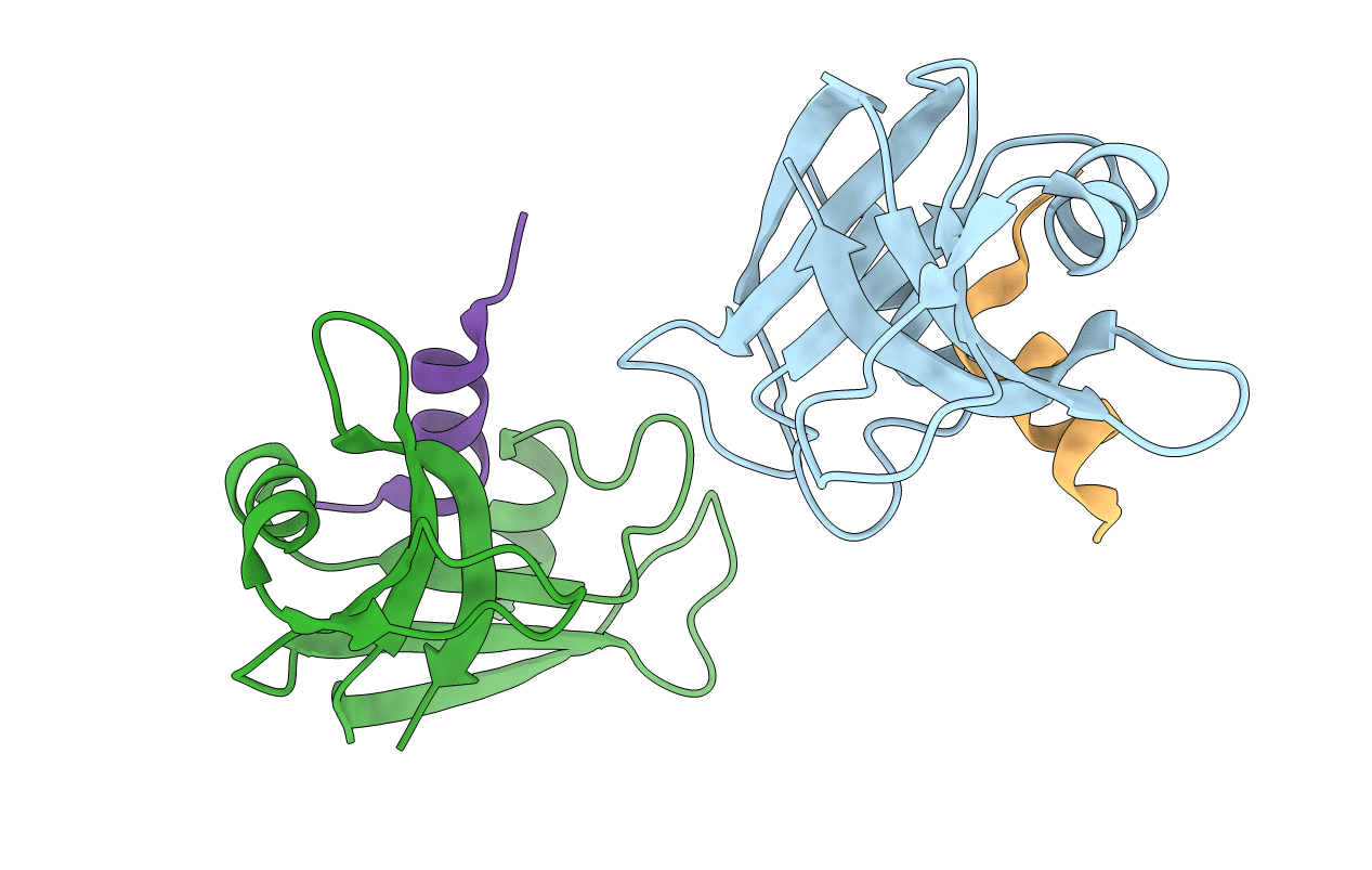
3OR0 image
Deposition Date 2010-09-05
Release Date 2010-10-20
Last Version Date 2024-11-06
Entry Detail
PDB ID:
3OR0
Keywords:
Title:
Semi-synthetic ribonuclease S: cyanylated homocysteine at position 13
Biological Source:
Source Organism:
Bos taurus (Taxon ID: 9913)
Method Details:
Experimental Method:
Resolution:
2.30 Å
R-Value Free:
0.27
R-Value Work:
0.21
R-Value Observed:
0.22
Space Group:
C 1 2 1
Macromolecular Entities
Polymer Type:polypeptide(L)
Molecule:Ribonuclease pancreatic
Gene (Uniprot):RNASE1
Chain IDs:B (auth: A), D (auth: B)
Chain Length:15
Number of Molecules:2
Biological Source:Bos taurus
Polymer Type:polypeptide(L)
Molecule:Ribonuclease pancreatic
Gene (Uniprot):RNASE1
Chain IDs:A (auth: a), C (auth: b)
Chain Length:15
Number of Molecules:2
Biological Source:Bos taurus
Modified Residue
Compound ID Chain ID Parent Comp ID Details 2D Image
4CY A MET NITRILO-L-METHIONINE
Primary Citation
Nitrile bonds as infrared probes of electrostatics in ribonuclease S.
J.Phys.Chem.B 114 13536 13544 (2010)
PMID: 20883003 DOI: 10.1021/jp106406p

Abstact

Three different nitrile-containing amino acids, p-cyanophenylalanine, m-cyanophenylalanine, and S-cyanohomocysteine, have been introduced near the active site of the semisynthetic enzyme ribonuclease S (RNase S) to serve as probes of electrostatic fields. Vibrational Stark spectra, measured directly on the probe-modified proteins, confirm the predominance of the linear Stark tuning rate in describing the sensitivity of the nitrile stretch to external electric fields, a necessary property for interpreting observed frequency shifts as a quantitative measure of local electric fields that can be compared with simulations. The X-ray structures of these nitrile-modified RNase variants and enzymatic assays demonstrate minimal perturbation to the structure and function, respectively, by the probes and provide a context for understanding the influence of the environment on the nitrile stretching frequency. We examine the ability of simulation techniques to recapitulate the spectroscopic properties of these nitriles as a means to directly test a computational electrostatic model for proteins, specifically that in the ubiquitous Amber-99 force field. Although qualitative agreement between theory and experiment is observed for the largest shifts, substantial discrepancies are observed in some cases, highlighting the ongoing need for experimental metrics to inform the development of theoretical models of electrostatic fields in proteins.

Legend

Protein

Chemical

Disease

Primary Citation of related structures