
Deposition Date
2010-09-01
Release Date
2011-04-27
Last Version Date
2024-02-21
Entry Detail
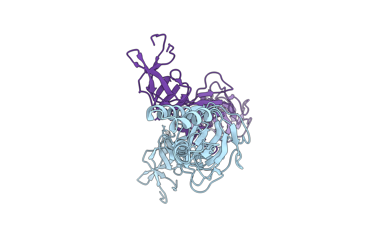
PDB ID:
3OPO
Keywords:
Title:
Crystal structure of the membrane fusion protein CusB from Escherichia coli
Biological Source:
Source Organism(s):
Escherichia coli K-12 (Taxon ID: 83333)
Expression System(s):
Method Details:
Experimental Method:
Resolution:
3.85 Å
R-Value Free:
0.33
R-Value Work:
0.29
R-Value Observed:
0.29
Space Group:
I 2 2 2
