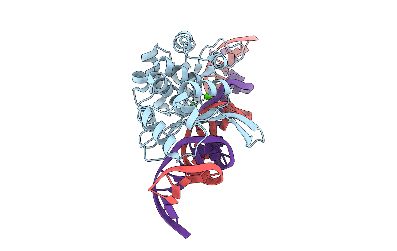
3OOL image
Entry Detail
PDB ID:
3OOL
Keywords:
Title:
I-SceI complexed with C/G+4 DNA substrate
Biological Source:
Source Organism:
Host Organism:
PDB Version:
Deposition Date:
2010-08-31
Release Date:
2010-11-17
Method Details:
Experimental Method:
Resolution:
2.30 Å
R-Value Free:
0.29
R-Value Work:
0.24
R-Value Observed:
0.24
Space Group:
C 1 2 1
Macromolecular Entities
Polymer Type:polypeptide(L)
Description:Intron encoded endonuclease I-SceI
Chain IDs:A
Chain Length:237
Number of Molecules:1
Biological Source:Saccharomyces cerevisiae
Ligand Molecules
Primary Citation
Evolution of I-SceI Homing Endonucleases with Increased DNA Recognition Site Specificity.
J.Mol.Biol. 405 185 200 (2011)
PMID: 21029741 DOI: 10.1016/j.jmb.2010.10.029

Abstact

Elucidating how homing endonucleases undergo changes in recognition site specificity will facilitate efforts to engineer proteins for gene therapy applications. I-SceI is a monomeric homing endonuclease that recognizes and cleaves within an 18-bp target. It tolerates limited degeneracy in its target sequence, including substitution of a C:G(+4) base pair for the wild-type A:T(+4) base pair. Libraries encoding randomized amino acids at I-SceI residue positions that contact or are proximal to A:T(+4) were used in conjunction with a bacterial one-hybrid system to select I-SceI derivatives that bind to recognition sites containing either the A:T(+4) or the C:G(+4) base pairs. As expected, isolates encoding wild-type residues at the randomized positions were selected using either target sequence. All I-SceI proteins isolated using the C:G(+4) recognition site included small side-chain substitutions at G100 and either contained (K86R/G100T, K86R/G100S and K86R/G100C) or lacked (G100A, G100T) a K86R substitution. Interestingly, the binding affinities of the selected variants for the wild-type A:T(+4) target are 4- to 11-fold lower than that of wild-type I-SceI, whereas those for the C:G(+4) target are similar. The increased specificity of the mutant proteins is also evident in binding experiments in vivo. These differences in binding affinities account for the observed ∼36-fold difference in target preference between the K86R/G100T and wild-type proteins in DNA cleavage assays. An X-ray crystal structure of the K86R/G100T mutant protein bound to a DNA duplex containing the C:G(+4) substitution suggests how sequence specificity of a homing enzyme can increase. This biochemical and structural analysis defines one pathway by which site specificity is augmented for a homing endonuclease.

Legend

Protein

Chemical

Disease

Primary Citation of related structures