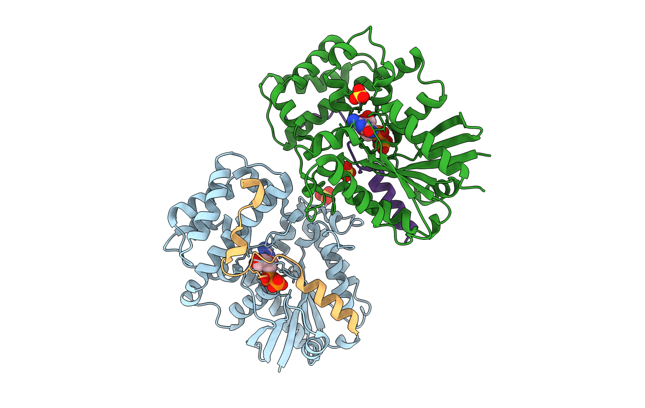
3ONW image
Deposition Date 2010-08-30
Release Date 2010-11-24
Last Version Date 2023-09-06
Entry Detail
PDB ID:
3ONW
Title:
Structure of a G-alpha-i1 mutant with enhanced affinity for the RGS14 GoLoco motif.
Biological Source:
Source Organism:
Homo sapiens (Taxon ID: 9606)
(Taxon ID: )
Host Organism:
Method Details:
Experimental Method:
Resolution:
2.38 Å
R-Value Free:
0.26
R-Value Work:
0.22
R-Value Observed:
0.22
Space Group:
P 2 2 21
Macromolecular Entities
Polymer Type:polypeptide(L)
Molecule:Guanine nucleotide-binding protein G(i) subunit alpha-1
Gene (Uniprot):GNAI1
Mutations:Q147L
Chain IDs:A, B
Chain Length:328
Number of Molecules:2
Biological Source:Homo sapiens
Polymer Type:polypeptide(L)
Molecule:Regulator of G-protein signaling 14
Gene (Uniprot):RGS14
Chain IDs:C, D
Chain Length:36
Number of Molecules:2
Biological Source:
Primary Citation
Structural Determinants of Affinity Enhancement between GoLoco Motifs and G-Protein {alpha} Subunit Mutants.
J.Biol.Chem. 286 3351 3358 (2011)
PMID: 21115486 DOI: 10.1074/jbc.M110.190496

Abstact

GoLoco motif proteins bind to the inhibitory G(i) subclass of G-protein α subunits and slow the release of bound GDP; this interaction is considered critical to asymmetric cell division and neuro-epithelium and epithelial progenitor differentiation. To provide protein tools for interrogating the precise cellular role(s) of GoLoco motif/Gα(i) complexes, we have employed structure-based protein design strategies to predict gain-of-function mutations that increase GoLoco motif binding affinity. Here, we describe fluorescence polarization and isothermal titration calorimetry measurements showing three predicted Gα(i1) point mutations, E116L, Q147L, and E245L; each increases affinity for multiple GoLoco motifs. A component of this affinity enhancement results from a decreased rate of dissociation between the Gα mutants and GoLoco motifs. For Gα(i1)(Q147L), affinity enhancement was seen to be driven by favorable changes in binding enthalpy, despite reduced contributions from binding entropy. The crystal structure of Gα(i1)(Q147L) bound to the RGS14 GoLoco motif revealed disorder among three peptide residues surrounding a well defined Leu-147 side chain. Monte Carlo simulations of the peptide in this region showed a sampling of multiple backbone conformations in contrast to the wild-type complex. We conclude that mutation of Glu-147 to leucine creates a hydrophobic surface favorably buried upon GoLoco peptide binding, yet the hydrophobic Leu-147 also promotes flexibility among residues 511-513 of the RGS14 GoLoco peptide.

Legend

Protein

Chemical

Disease

Primary Citation of related structures