3ONM image
Deposition Date 2010-08-30
Release Date 2011-01-26
Last Version Date 2024-02-21
Entry Detail
PDB ID:
3ONM
Keywords:
Title:
Effector binding Domain of LysR-Type transcription factor RovM from Y. pseudotuberculosis
Biological Source:
Source Organism:
Host Organism:
Method Details:
Experimental Method:
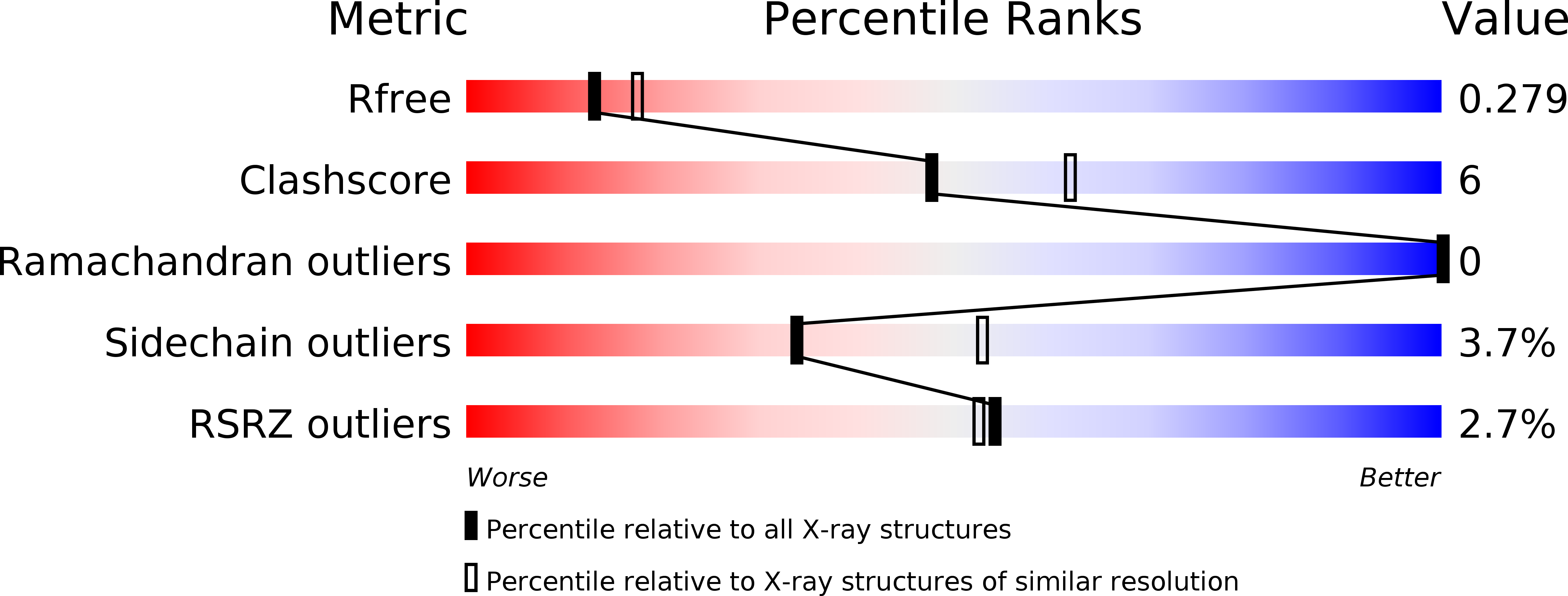
Resolution:
2.40 Å
R-Value Free:
0.29
R-Value Work:
0.22
R-Value Observed:
0.22
Space Group:
I 41 2 2
Macromolecular Entities
Polymer Type:polypeptide(L)
Molecule:Transcriptional regulator LrhA
Chain IDs:A, B
Chain Length:238
Number of Molecules:2
Biological Source:Yersinia pseudotuberculosis
Primary Citation
Structure of the effector-binding domain of the LysR-type transcription factor RovM from Yersinia pseudotuberculosis.
Acta Crystallogr.,Sect.D 67 81 90 (2011)
PMID: 21245528 DOI: 10.1107/S0907444910049681

Abstact

In enteropathogenic Yersinia, the expression of several early-phase virulence factors such as invasin is tightly regulated in response to environmental cues. The responsible regulatory network is complex, involving several regulatory RNAs and proteins such as the LysR-type transcription regulator (LTTR) RovM. In this study, the crystal structure of the effector-binding domain (EBD) of RovM, the first LTTR protein described as being involved in virulence regulation, was determined at a resolution of 2.4 Å. Size-exclusion chromatography and comparison with structures of full-length LTTRs show that RovM is most likely to adopt a tetrameric arrangement with two distant DNA-binding domains (DBDs), causing the DNA to bend around it. Additionally, a cavity was detected in RovM which could bind small inducer molecules.

Legend

Protein

Chemical

Disease

Primary Citation of related structures