3OMC image
Entry Detail
PDB ID:
3OMC
Title:
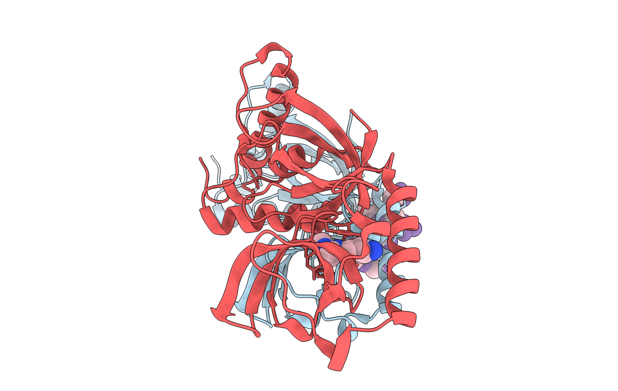
Structure of human SND1 extended tudor domain in complex with the symmetrically dimethylated arginine PIWIL1 peptide R4me2s
Biological Source:
Host Organism:
PDB Version:
Deposition Date:
2010-08-26
Release Date:
2010-09-15
Method Details:
Experimental Method:
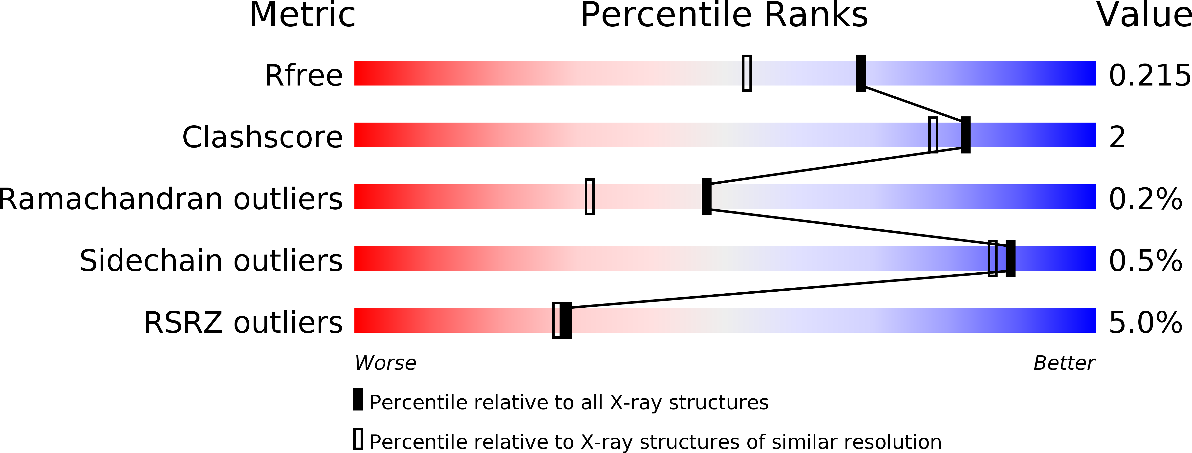
Resolution:
1.77 Å
R-Value Free:
0.21
R-Value Work:
0.17
R-Value Observed:
0.18
Space Group:
P 1 21 1
Macromolecular Entities
Polymer Type:polypeptide(L)
Description:Staphylococcal nuclease domain-containing protein 1
Chain IDs:A, B
Chain Length:261
Number of Molecules:2
Biological Source:Homo sapiens
Polymer Type:polypeptide(L)
Description:SYNTHETIC PEPTIDE
Chain IDs:C, D
Chain Length:8
Number of Molecules:2
Biological Source:synthetic construct
Modified Residue
Compound ID Chain ID Parent Comp ID Details 2D Image
2MR C ARG N3, N4-DIMETHYLARGININE
Ligand Molecules
Primary Citation
Structural basis for recognition of arginine methylated Piwi proteins by the extended Tudor domain.
Proc.Natl.Acad.Sci.USA 107 18398 18403 (2010)
PMID: 20937909 DOI: 10.1073/pnas.1013106107

Abstact

Arginine methylation modulates diverse cellular processes and represents a molecular signature of germ-line-specific Piwi family proteins. A subset of Tudor domains recognize arginine methylation modifications, but the binding mechanism has been lacking. Here we establish that, like other germ-line Tudor proteins, the ancestral staphylococcal nuclease domain-containing 1 (SND1) polypeptide is expressed and associates with PIWIL1/Miwi in germ cells. We find that human SND1 binds PIWIL1 in an arginine methylation-dependent manner with a preference for symmetrically dimethylated arginine. The entire Tudor domain and a bifurcated SN domain are required for this binding activity, whereas the canonical Tudor domain alone is insufficient for methylarginine ligand binding. Crystal structures show that the intact SND1 extended Tudor domain forms a wide and negatively charged binding groove, which can accommodate distinct symmetrically dimethylated arginine peptides from PIWIL1 in different orientations. This analysis explains how SND1 preferentially recognizes symmetrical dimethylarginine via an aromatic cage and conserved hydrogen bonds, and provides a general paradigm for the binding mechanisms of methylarginine-containing peptides by extended Tudor domains.

Legend

Protein

Chemical

Disease

Primary Citation of related structures