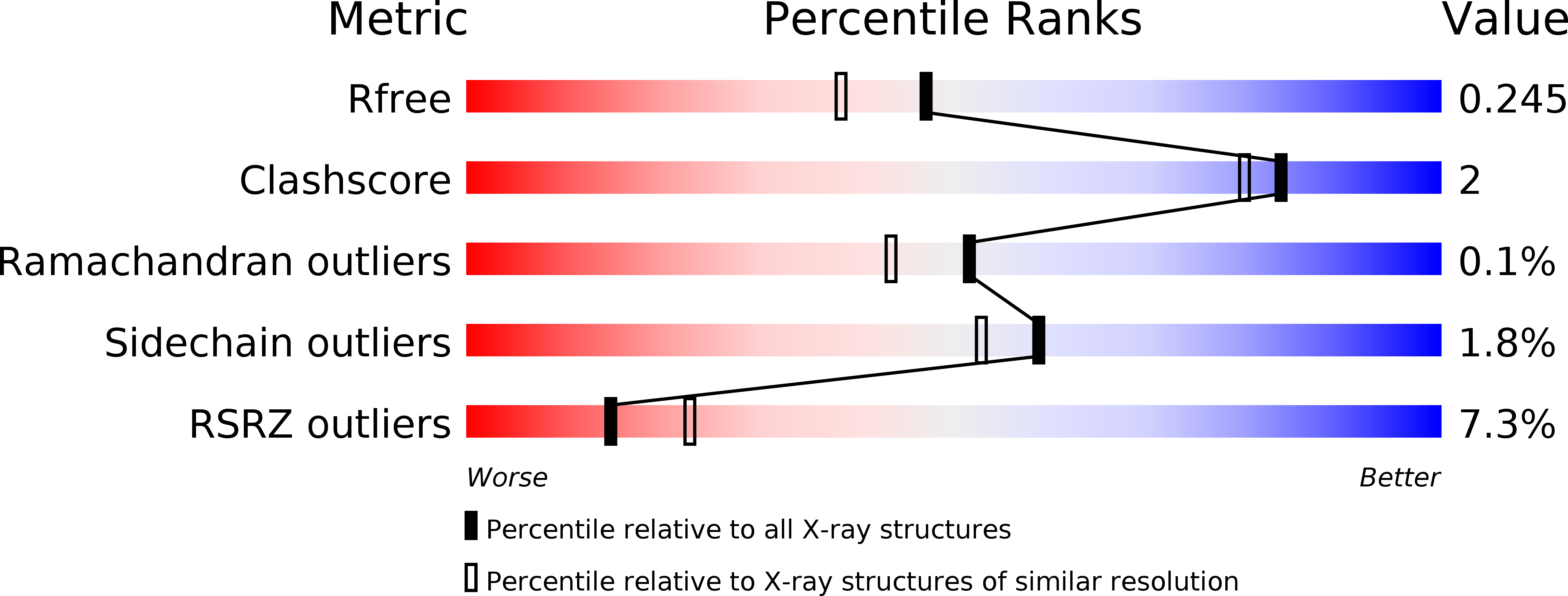
3OM6 image
Deposition Date 2010-08-26
Release Date 2011-03-23
Last Version Date 2024-02-21
Entry Detail
PDB ID:
3OM6
Keywords:
Title:
Crystal structure of B. megaterium levansucrase mutant Y247A
Biological Source:
Source Organism:
Host Organism:
Method Details:
Experimental Method:
Resolution:
1.96 Å
R-Value Free:
0.22
R-Value Work:
0.22
R-Value Observed:
0.22
Space Group:
P 1 21 1
Macromolecular Entities
Polymer Type:polypeptide(L)
Molecule:Levansucrase
Gene (Uniprot):sacB
Mutations:Y247A
Chain IDs:A, B, C, D
Chain Length:456
Number of Molecules:4
Biological Source:Bacillus megaterium
Primary Citation
Polysaccharide Synthesis of the Levansucrase SacB from Bacillus megaterium Is Controlled by Distinct Surface Motifs.
J.Biol.Chem. 286 17593 17600 (2011)
PMID: 21454585 DOI: 10.1074/jbc.M110.203166

Abstact

Despite the widespread biological function of carbohydrates, the polysaccharide synthesis mechanisms of glycosyltransferases remain largely unexplored. Bacterial levansucrases (glycoside hydrolase family 68) synthesize high molecular weight, β-(2,6)-linked levan from sucrose by transfer of fructosyl units. The kinetic and biochemical characterization of Bacillus megaterium levansucrase SacB variants Y247A, Y247W, N252A, D257A, and K373A reveal novel surface motifs remote from the sucrose binding site with distinct influence on the polysaccharide product spectrum. The wild type activity (k(cat)) and substrate affinity (K(m)) are maintained. The structures of the SacB variants reveal clearly distinguishable subsites for polysaccharide synthesis as well as an intact active site architecture. These results lead to a new understanding of polysaccharide synthesis mechanisms. The identified surface motifs are discussed in the context of related glycosyltransferases.

Legend

Protein

Chemical

Disease

Primary Citation of related structures