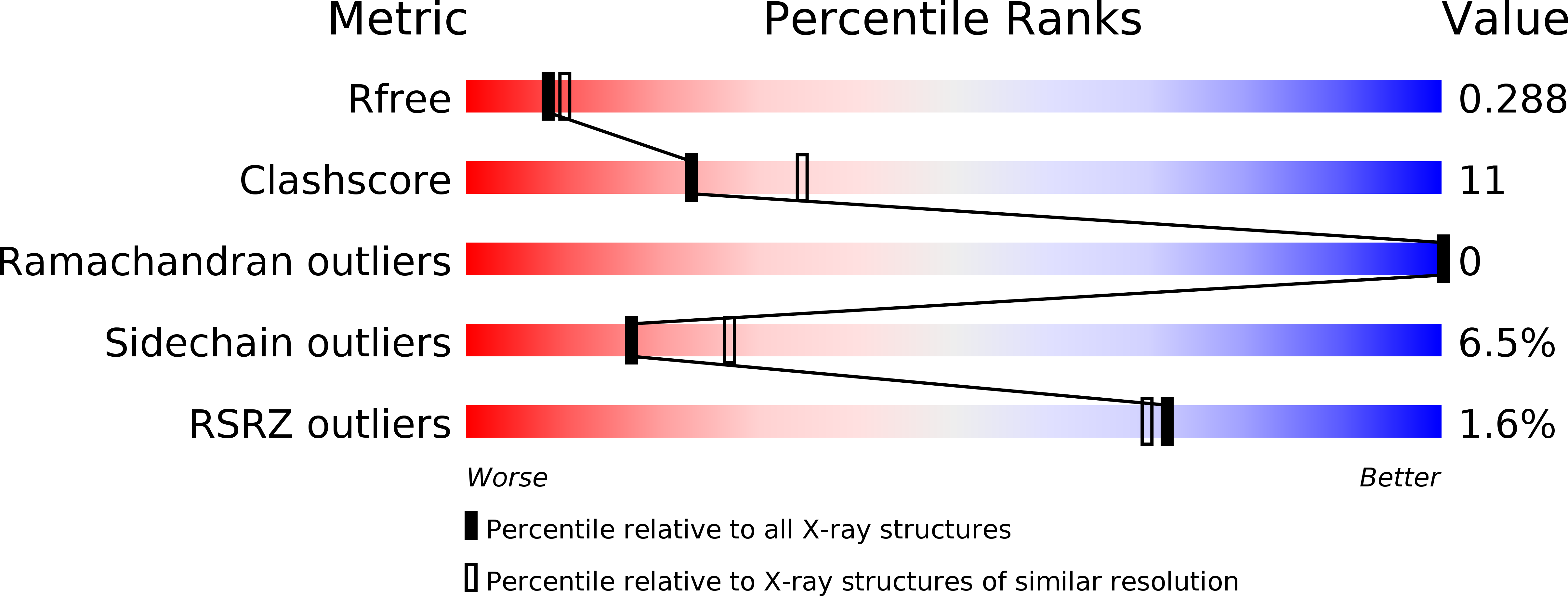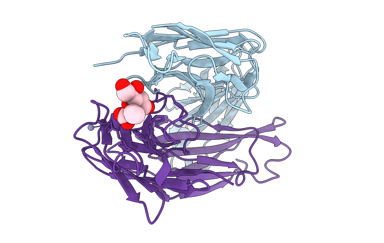
3OKE image
Deposition Date 2010-08-24
Release Date 2011-04-06
Last Version Date 2024-10-30
Entry Detail
PDB ID:
3OKE
Keywords:
Title:
Crystal structure of S25-39 in complex with Ko
Biological Source:
Source Organism:
Mus musculus (Taxon ID: 10090)
Method Details:
Experimental Method:
Resolution:
2.40 Å
R-Value Free:
0.28
R-Value Work:
0.22
R-Value Observed:
0.22
Space Group:
P 21 21 21
Macromolecular Entities
Polymer Type:polypeptide(L)
Molecule:S25-39 Fab (IgG1k) light chain
Chain IDs:A
Chain Length:219
Number of Molecules:1
Biological Source:Mus musculus
Polymer Type:polypeptide(L)
Molecule:S25-39 Fab (IgG1k) heavy chain
Chain IDs:B
Chain Length:222
Number of Molecules:1
Biological Source:Mus musculus
Primary Citation
A Common NH53K Mutation in the Combining Site of Antibodies Raised against Chlamydial LPS Glycoconjugates Significantly Increases Avidity.
Biochemistry 50 3357 3368 (2011)
PMID: 21405106 DOI: 10.1021/bi101886v

Abstact

The crystal structures of the antigen-binding fragment of the murine monoclonal antibody (mAb) S25-39 in the presence of several antigens representing chlamydial lipopolysaccharide (LPS) epitopes based on the bacterial sugar 3-deoxy-α-D-manno-oct-2-ulosonic acid (Kdo) have been determined at resolutions from 2.4 to 1.8 Å. The antigen-binding site of this antibody differs from the well-characterized antibody S25-2 by a single mutation away from the germline of asparagine H53 to lysine, yet this one mutation results in a significant increase in avidity across a range of antigens. A comparison of the two antibody structures reveals that the mutated Lys H53 forms additional hydrogen bonds and/or charged-residue interactions with the second Kdo residue of every antigen having two or more carbohydrate residues. Significantly, the NH53K mutation results from a single nucleotide substitution in the germline sequence common among a panel of antibodies raised against glycoconjugates containing carbohydrate epitopes of chlamydial LPS. Like S25-2, S25-39 displays significant induced fit of complementarity determining region (CDR) H3 upon antigen binding, with the unliganded structure possessing a conformation distinct from those reported earlier for S25-2. The four different observed conformations for CDR H3 suggest that this CDR has evolved to exploit the recognition potential of a flexible loop while minimizing the associated entropic penalties of binding by adopting a limited number of ordered conformations in the unliganded state. These observations reveal strategies evolved to balance adaptability and specificity in the germline antibody response to carbohydrate antigens.

Legend

Protein

Chemical

Disease

Primary Citation of related structures