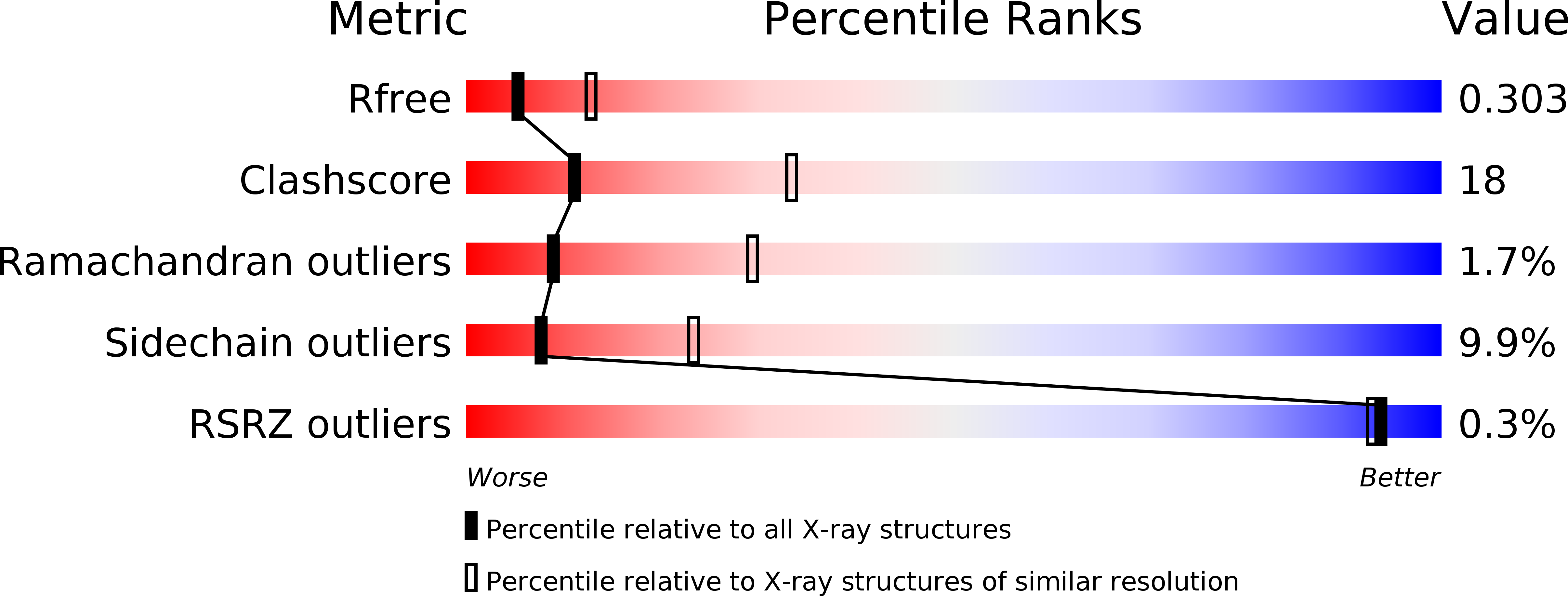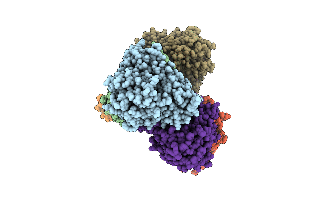
3OFU image
Deposition Date 2010-08-16
Release Date 2011-05-04
Last Version Date 2023-11-01
Entry Detail
PDB ID:
3OFU
Keywords:
Title:
Crystal Structure of Cytochrome P450 CYP101C1
Biological Source:
Source Organism:
Host Organism:
Method Details:
Experimental Method:
Resolution:
2.80 Å
R-Value Free:
0.30
R-Value Work:
0.19
R-Value Observed:
0.19
Space Group:
P 1 21 1
Macromolecular Entities
Structures with similar UniProt ID
Protein Blast
Polymer Type:polypeptide(L)
Molecule:Cytochrome P450
Gene (Uniprot):Saro_2249
Chain IDs:A, B, C, D, E, F
Chain Length:396
Number of Molecules:6
Biological Source:Novosphingobium aromaticivorans
Primary Citation
Structural Analysis of CYP101C1 from Novosphingobium aromaticivorans DSM12444.
Chembiochem 12 88 99 (2011)
PMID: 21154803 DOI: 10.1002/cbic.201000537

Abstact

CYP101C1 from Novosphingobium aromaticivorans DSM12444 is a homologue of CYP101D1 and CYP101D2 enzymes from the same bacterium and CYP101A1 from Pseudomonas putida. CYP101C1 does not bind camphor but is capable of binding and hydroxylating ionone derivatives including α- and β-ionone and β-damascone. The activity of CYP101C1 was highest with β-damascone (k(cat)=86 s(-1)) but α-ionone oxidation was the most regioselective (98 % at C3). The crystal structures of hexane-2,5-diol- and β-ionone-bound CYP101C1 have been solved; both have open conformations and the hexanediol-bound form has a clear access channel from the heme to the bulk solvent. The entrance of this channel is blocked when β-ionone binds to the enzyme. The heme moiety of CYP101C1 is in a significantly different environment compared to the other structurally characterised CYP101 enzymes. The likely ferredoxin binding site on the proximal face of CYP101C1 has a different topology but a similar overall positive charge compared to CYP101D1 and CYP101D2, all of which accept electrons from the ArR/Arx class I electron transfer system.

Legend

Protein

Chemical

Disease

Primary Citation of related structures