Deposition Date
2010-08-14
Release Date
2010-12-08
Last Version Date
2024-11-06
Entry Detail
PDB ID:
3OF9
Keywords:
Title:
Structural Basis for Irreversible Inhibition of Human Cathepsin L by a Diazomethylketone Inhibitor
Biological Source:
Source Organism(s):
Homo sapiens (Taxon ID: 9606)
Expression System(s):
Method Details:
Experimental Method:
Resolution:
1.76 Å
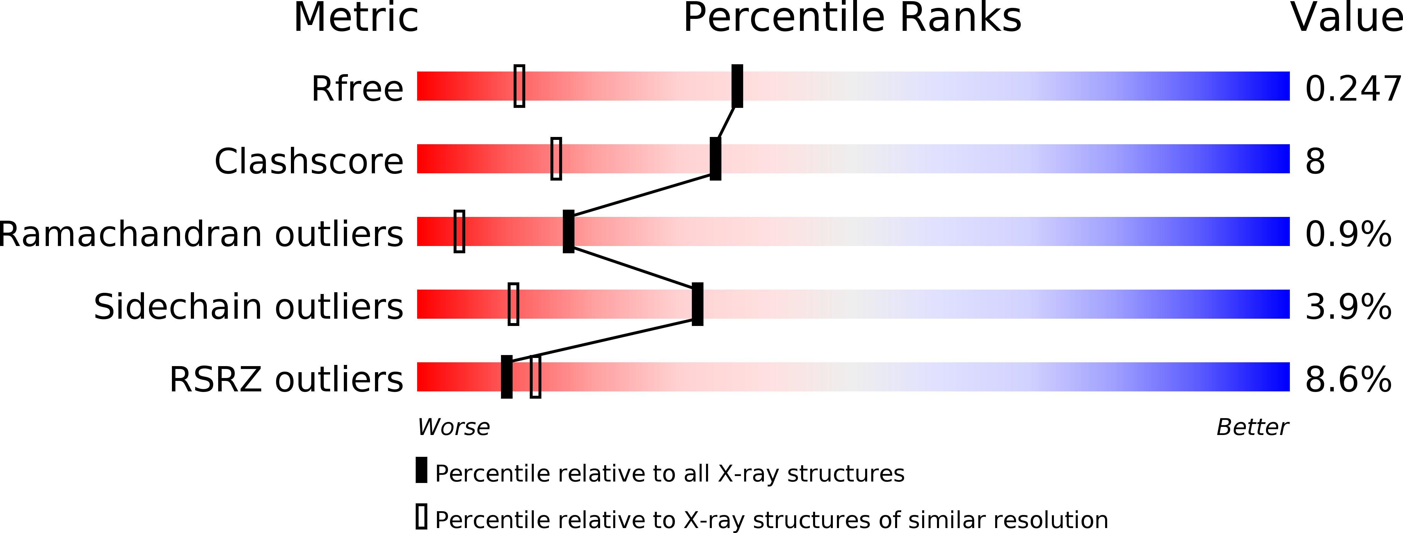
R-Value Free:
0.24
R-Value Work:
0.22
R-Value Observed:
0.22
Space Group:
P 65