Deposition Date
2010-08-12
Release Date
2010-09-15
Last Version Date
2023-09-06
Entry Detail
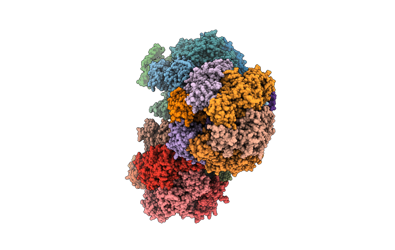
PDB ID:
3OE7
Keywords:
Title:
Structure of four mutant forms of yeast f1 ATPase: gamma-I270T
Biological Source:
Source Organism(s):
Saccharomyces cerevisiae (Taxon ID: 4932)
Expression System(s):
Method Details:
Experimental Method:
Resolution:
3.19 Å
R-Value Free:
0.24
R-Value Work:
0.18
R-Value Observed:
0.19
Space Group:
P 1 21 1