3ODQ image
Deposition Date 2010-08-11
Release Date 2010-12-01
Last Version Date 2023-09-06
Entry Detail
PDB ID:
3ODQ
Title:
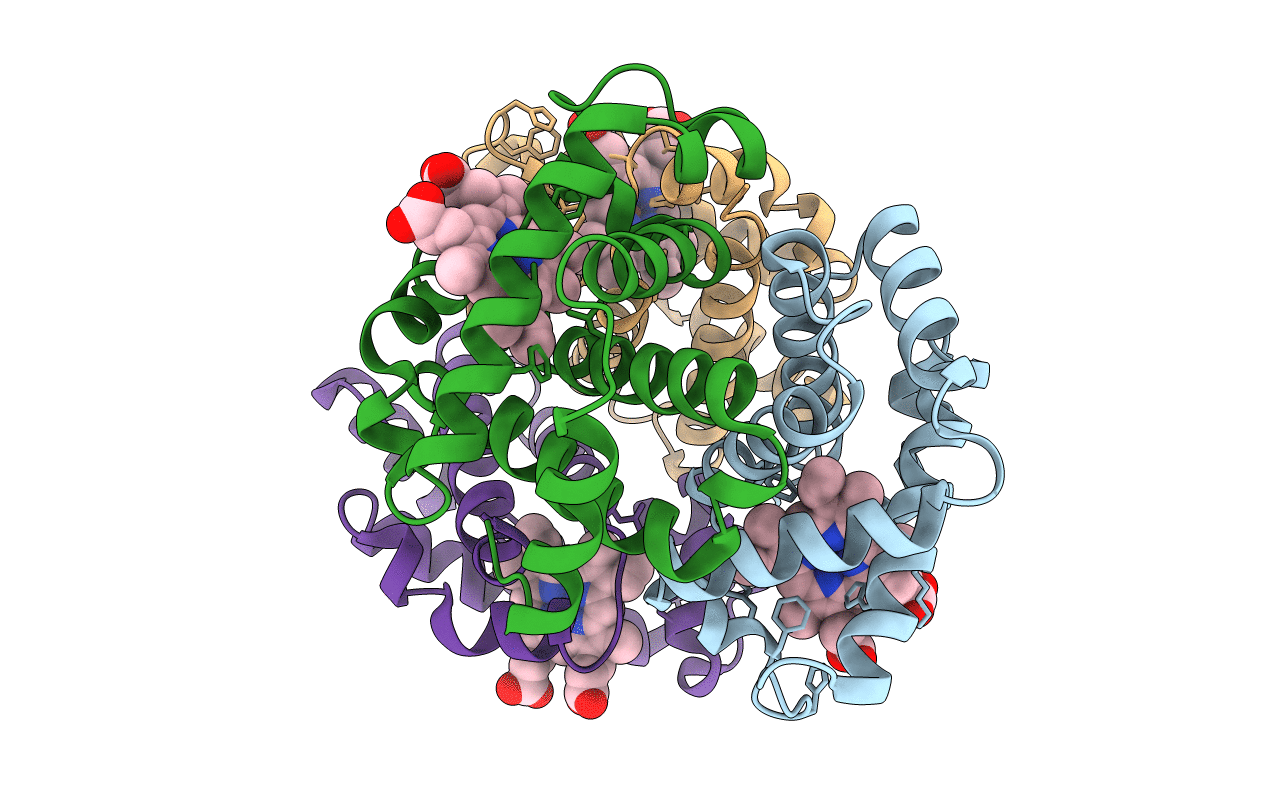
Structure of a Crystal Form of Human Methemoglobin Indicative of Fiber Formation
Biological Source:
Source Organism:
Homo sapiens (Taxon ID: 9606)
Method Details:
Experimental Method:
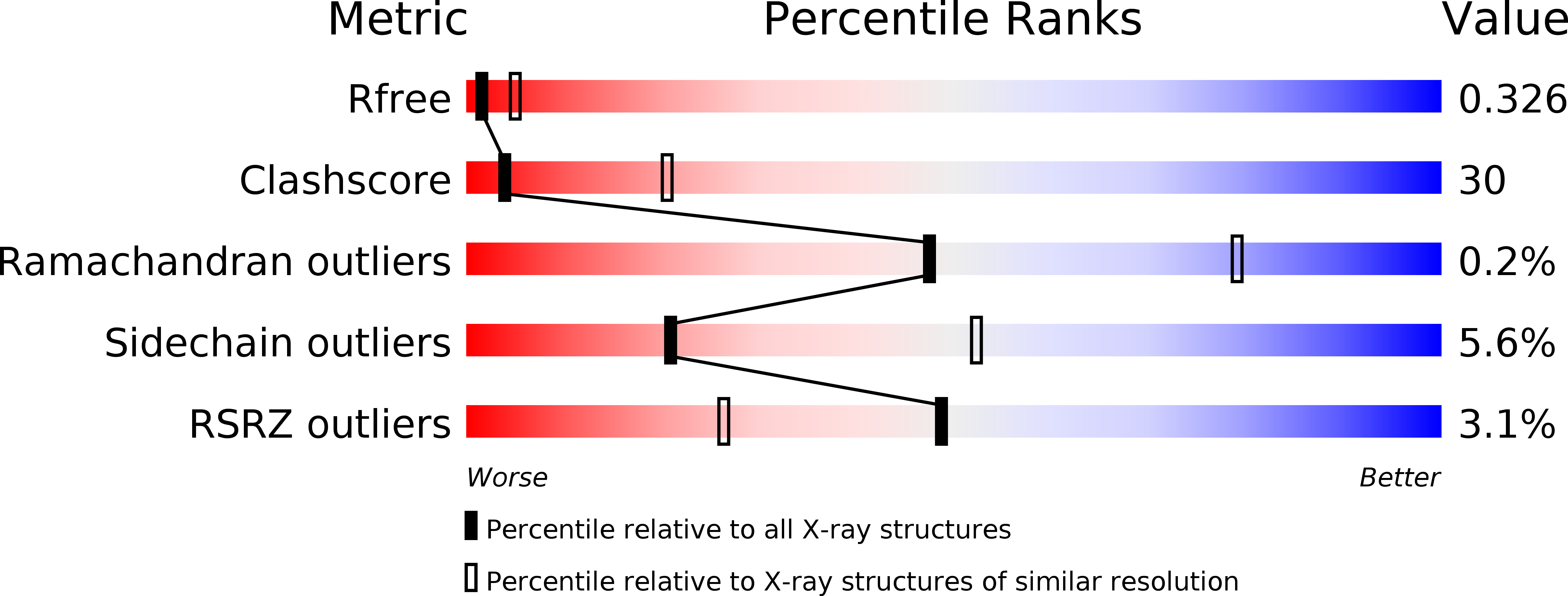
Resolution:
3.10 Å
R-Value Free:
0.32
R-Value Work:
0.28
R-Value Observed:
0.28
Space Group:
P 61 2 2
Macromolecular Entities
Polymer Type:polypeptide(L)
Molecule:Hemoglobin subunit alpha
Gene (Uniprot):HBA1, HBA2
Chain IDs:A, C
Chain Length:141
Number of Molecules:2
Biological Source:Homo sapiens
Polymer Type:polypeptide(L)
Molecule:Hemoglobin subunit beta
Gene (Uniprot):HBB
Chain IDs:B, D
Chain Length:146
Number of Molecules:2
Biological Source:Homo sapiens
Ligand Molecules
Primary Citation
Structure of a crystal form of human methemoglobin indicative of fiber formation.
Acta Crystallogr.,Sect.D 66 1316 1322 (2010)
PMID: 21123872 DOI: 10.1107/S0907444910040370

Abstact

Human methemoglobin was crystallized in a unique unit cell and its structure was solved by molecular replacement. The hexagonal unit cell has unit-cell parameters a = b = 54.6, c = 677.4 Å, with symmetry consistent with space group P6₁22. The unit cell has the second highest aspect ratio of all unit cells contained in the PDB. The 12 molecules in the unit cell describe a right-handed helical filament having no polarity, which is different from the filament composed of HbS fibers, which is the only other well characterized fiber of human hemoglobin. The filaments reported here can be related to canonical sickle-cell hemoglobin filaments and to an alternative sickle-cell filament deduced from fiber diffraction by slight modifications of intermolecular contacts.

Legend

Protein

Chemical

Disease

Primary Citation of related structures