3OD9 image
Deposition Date 2010-08-11
Release Date 2010-12-22
Last Version Date 2024-02-21
Entry Detail
PDB ID:
3OD9
Title:
Crystal structure of PliI-Ah, periplasmic lysozyme inhibitor of I-type lysozyme from Aeromonas hydrophyla
Biological Source:
Source Organism:
Host Organism:
Method Details:
Experimental Method:
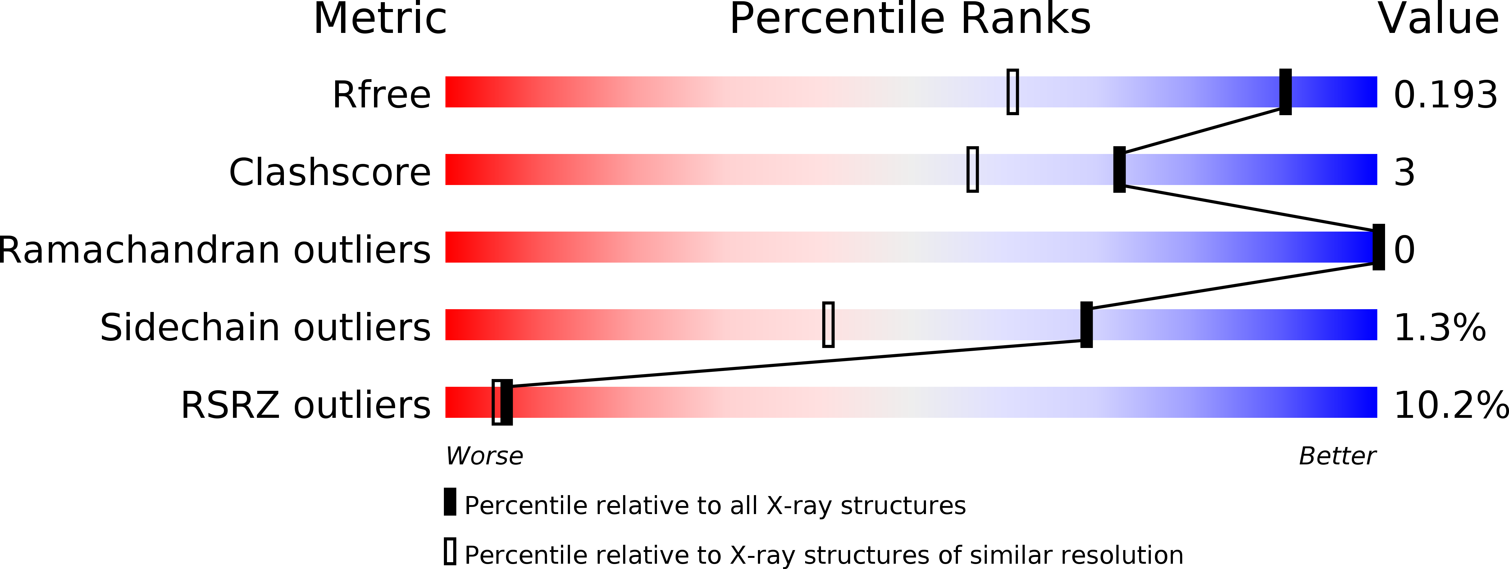
Resolution:
1.41 Å
R-Value Free:
0.19
R-Value Work:
0.16
R-Value Observed:
0.16
Space Group:
C 1 2 1
Macromolecular Entities
Polymer Type:polypeptide(L)
Molecule:Putative exported protein
Gene (Uniprot):AHA_1205
Chain IDs:A, B, C
Chain Length:135
Number of Molecules:3
Biological Source:Aeromonas hydrophila
Primary Citation
Molecular Basis of Bacterial Defense against Host Lysozymes: X-ray Structures of Periplasmic Lysozyme Inhibitors PliI and PliC.
J.Mol.Biol. 405 1233 1245 (2011)
PMID: 21146533 DOI: 10.1016/j.jmb.2010.12.007

Abstact

Lysozymes play a key role in the innate immune system of vertebrates and invertebrates by hydrolyzing peptidoglycan, a vital component of the bacterial cell wall. Gram-negative bacteria produce various types of lysozyme inhibitors that allow them to survive the bactericidal action of lysozyme when their outer membrane is permeabilized. So far, three lysozyme inhibitor families have been described: the Ivy (inhibitor of vertebrate lysozyme) family, the MliC/PliC (membrane-associated/periplasmic lysozyme inhibitor of C-type lysozyme) family, and the PliI (periplasmic lysozyme inhibitor of I-type lysozyme) family. Here, we report high-resolution crystal structures of Salmonella typhimurium PliC (PliC-St) and Aeromonas hydrophila PliI (PliI-Ah). The structure of PliI-Ah is the first in the recently discovered PliI family of lysozyme inhibitors, while the structure of PliC-St is the first structure of a periplasmic lysozyme inhibitor from the PliC/MliC family. Using small-angle X-ray scattering, we demonstrate that both PliC-St and PliI-Ah form stable dimers in solution. The functional dimer architecture of PliC-St is very different from that of the recently described MliC from Pseudomonas aeruginosa (MliC-Pa), despite the close resemblance of their monomers. Furthermore, PliI-Ah has distinctly different monomer and dimer folds compared to PliC, MliC, and Ivy proteins. Site-directed mutagenesis suggests that the inhibitory action of PliI-Ah proceeds via an insertion of a loop containing the conserved SGxY motif into the active center of I-type lysozymes. This motif is related to the functional SGxxY motif found in the MliC/PliC family.

Legend

Protein

Chemical

Disease

Primary Citation of related structures