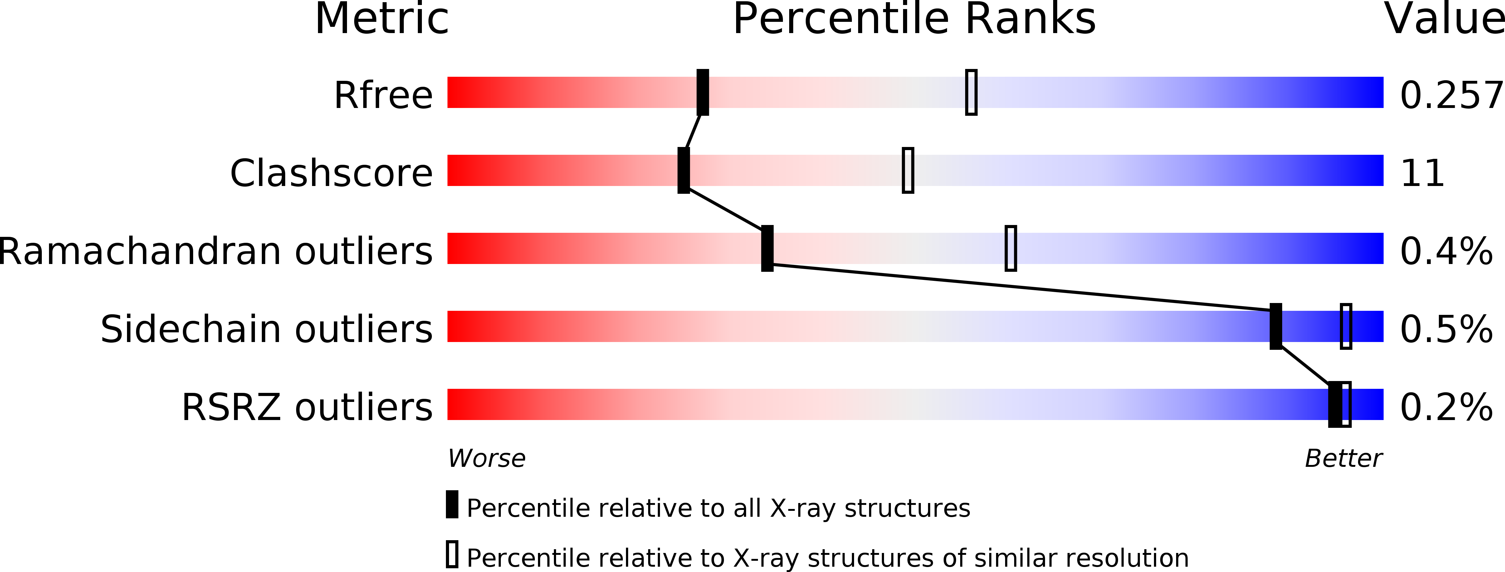
3OB4 image
Deposition Date 2010-08-06
Release Date 2011-02-02
Last Version Date 2024-10-09
Entry Detail
PDB ID:
3OB4
Keywords:
Title:
MBP-fusion protein of the major peanut allergen Ara h 2
Biological Source:
Source Organism:
Escherichia coli (Taxon ID: 562)
Arachis hypogaea (Taxon ID: 3818)
Host Organism:
Method Details:
Experimental Method:
Resolution:
2.71 Å
R-Value Free:
0.25
R-Value Work:
0.18
R-Value Observed:
0.18
Space Group:
C 1 2 1
Macromolecular Entities
Polymer Type:polypeptide(L)
Molecule:Maltose ABC transporter periplasmic protein,Arah 2
Mutations:D82A,K83A,E172A,N173A,K239A
Chain IDs:A
Chain Length:500
Number of Molecules:1
Biological Source:Escherichia coli, Arachis hypogaea
Peptide-like Molecules
PRD_900009
Primary Citation
Ara h 2: crystal structure and IgE binding distinguish two subpopulations of peanut allergic patients by epitope diversity.
Allergy 66 878 885 (2011)
PMID: 21255036 DOI: 10.1111/j.1398-9995.2010.02532.x

Abstact

BACKGROUND Peanut allergy affects 1% of the population and causes the most fatal food-related anaphylactic reactions. The protein Ara h 2 is the most potent peanut allergen recognized by 80-90% of peanut allergic patients. METHODS The crystal structure of the major peanut allergen Ara h 2 was determined for the first time at 2.7 Å resolution using a customized maltose-binding protein (MBP)-fusion system. IgE antibody binding to the MBP fusion construct vs the natural allergen was compared by ELISA using sera from peanut allergic patients. RESULTS The structure of Ara h 2 is a five-helix bundle held together by four disulfide bonds and related to the prolamin protein superfamily. The fold is most similar to other amylase and trypsin inhibitors. The MBP--Ara h 2 fusion construct was positively recognized by IgE from 76% of allergic patients (25/33). Two populations of patients could be identified. Subpopulation 1 (n = 14) showed an excellent correlation of IgE antibody binding to natural vs recombinant Ara h 2. Subpopulation 2 (n = 15) showed significantly reduced IgE binding to the MBP fusion protein. Interestingly, about 20% of the IgE binding in subpopulation 2 could be recovered by increasing the distance between MBP and Ara h 2 in a second construct. DISCUSSION The reduced IgE binding to the MBP--Ara h 2 of subpopulation 2 indicates that the MBP molecule protects an immunodominant epitope region near the first helix of Ara h 2. Residues involved in the epitope(s) are suggested by the crystal structure. The MBP--Ara h 2 fusion constructs will be useful to further elucidate the relevance of certain epitopes to peanut allergy.

Legend

Protein

Chemical

Disease

Primary Citation of related structures